PT/INRs: Helping Patients Manage their Anticoagulation

So, who’s up for some bloodwork? Venipuncture, anyone? Finger prick, maybe? No..? Oh… Well, ok then. I’ll carry on. I know it may sound a bit crazy, but as a PT, I actually do some (very) minor blood work. I’m not trained as a nurse or phlebotomist or anything like that. Nope. Just some on-the-job-training on doing PT/INRs.

In this case, PT doesn’t stand for Physical Therapist like it usually does in things I write and across the medical spectrum. In this case, PT stands for prothrombin time (or “protime”) and INR stands for international normalized ratio. These factors combine to tell us many things about the effectiveness fo anticoagulant therapy. And, I’m sure you guessed it, they are important for us, as rehab clinicians, to do our best work for our patients. A normal INR in a patient who is not pharmacologically anticoagulated is 1.0. When we are pharmacologically anticoagulating, we want the INR to be between 2.0 and 3.0 and I have even been asked to go up to 3.5 for certain patients with multiple risk factors or who have mechanical heart valves. Average PT is 10-14 seconds.

IF you find that INR is greater than 5.0, you should NOT be performing any kind of therapeutic intervention and you should NOT be exercising the patient. Obviously, they will need to move enough to access care, and they should do so safely, maybe even with your help to prevent falls, but outside of that, they are inappropriate for therapy. If you find an INR higher than 6.0, the patients needs to get immediate medical attention as they are at an unsafe risk of bleeding. If you find the INR is too low, mobilization helps with blood flow which decreases clotting, so you should be working to mobilize the patient as much as you can in the absence of signs and sympyoms of DVT or PEs. However, you will still need to contact the physician as the patient may need adjustments in their anticoagulant dosage.


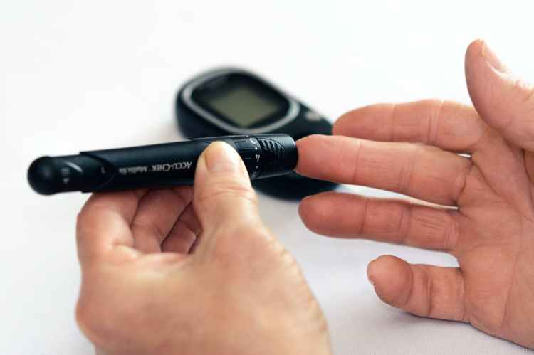
Doing this is actually really easy. We use a small meter called a CoaguCheck. Some patients have their own but I carried one regularly because I did so many of these. A small finger prick is done with clean technique, a capillary chip is inserted in to the machine, the blood is placed on the capillary chip, and a reading appears. There are several more pieces to this because the machines can be VERY finicky. The biggest issue is usually getting blood! I’ve put some serious time in to managing an arm from proximal to distal in an effort to “milk” blood to the finger to make sure I can get enough.

The best tip I ever received in performing this procedure is that you need enough blood to make it looks like a lady bug is sitting on the finger. This needs to be significantly more blood than what is required for blood glucose monitoring. I know that doesn’t sound like a lot, but if that blood is not therapeutically coagulated, it can be really difficult to get that much in the amount of time you are given. That’s the other hard part, the machine gives you a countdown in which you must apply the blood to the capillary chip. If you run the timer down, you have to start over with a new finger prick, new chip, and new reading.

Roche Diagnostics CoaguChek XS System Coaguchek XS System:Healthcare
Source

I typically go at this in a specific way each time: getting everything set up with clean technique, then get the chip, machine, and lancet ready. I then start my timer (which happens when you insert the capillary chip in to the meter) and spend about half the time (90 seconds) working the arm, hand, and finger to make sure I am going to get enough blood. Then I clean the finger, lance it, and “milk” enough blood out to look like a lady bug is sitting on the finger. Finally, I apply the capillary chip to the blood, allow capillary action to do its thing, and then wait for my reading.


Especially after a surgery, many of our patients are on prohpylactic anticoagulant therapies to reduce their risk of developing blood clots that could be potentially fatal if they travel throughout the circulatory system and relocate themselves in the lungs. But post-operative patients are not the only ones I help with this minor technique. As many of you know, I manage many patients with complex chronic diseases that require them to be heavily anticoagulated. These conditions may include heart failure with atrial fibrillation, cancer when chemo effects to the heart, or they have hypertension, diabetes, and Factor V Leiden deficiency. There are a million combinations I could list here. Regardless, most of them end up on Coumadin/Warfarin for anticoagulation which means their PT/INRs have to be monitored for therapeutic effect.

Now, of course there are other options for long term anticoagulation, such a rivaroxaban (Xarelto) or apixaban (Eliquis). These have some serious benefits like not having your finger pricked weekly to monitor your PT/INR, not having to worry about dietary choices (see below), and having fewer drug-to-drug interactions. However, when you have a patient with a history or high risk of falls, you don’t really want them on these particular options. Even though the effects wear off sooner, once the anticoagulant is in effect, it cannot be reversed. Once you start bleeding, you keep bleeding. That’s a real concern when you develop a subdural hematoma from a fall.

Photo by Karolina Grabowska on Pexels.com

Another drug in this “new” class of anticoagulants is Dabigatran (Pradaxa). I listed this one separately because a clotting antidote has actually been developed called idarucizumab (PraxBind). However, Pradaxa can still be hard on kidney function and tends to be incompatible with mechanical heart valves, similar to the other drugs in the “new” class of anticoagulants.


Things to watch out for when you have a patient utilizing Coumadin/Warfarin (outside of the obvious bruising/bleeding) include:

  • Medication Interactions. All generally administered medications included
    • Products containing acetaminophen
    • Most broad spectrum antibiotics
    • Aspirin
    • all other NSAIDs
    • Most antacids and laxatives
    • Antifungals
    • Amiodarone (or other rhythm drugs)
  • Supplements. Just don’t. There are many interactions with various supplements and they are not well controlled or easy to treat.
  • Foods. Anything high in Vitamin K. This means all leafy green and dark purple vegetables and fruits. This also means green tea. Like I mentioned above, Vitamin K is the antidote to Coumadin bleeding, so eating foods with higher levels of vitamin K will decrease the effectiveness of the drug.
    • Other foods include grapefruit, cranberry, garlic, and black licorice
  • Alcohol. Alcohol is also a blood thinner so you don’t want to double down on the effects of the medications.

Also, be on the lookout for bleeding that isn’t obvious. This can be GI bleeding, subdermal bleeding, internal bleeding, or cranial bleeding which would result in:

black or coffee-grounds stool or vomit
bleeding of the gums when brushing teeth (not otherwise explained)
severe head aches or stomach aches
dizziness, weakness, fatigue
low HgB and low Hct on CBCs
shortness of breath with minimal activity or at rest
new onset or increase in falls
ecchymosis of an entire limb or quadrant

All of these things would warrant immediate evaluation by a medical physician, probably in urgent care or emergency depending on the severity. Patients may need to receive the Coumadin antidote, Vitamin K or PCC.


Photo by Daria Shevtsova on Pexels.com

Other considerations include the need for dietary support. Call in a consult to your dietician because these folks aren’t going to be able to eat much from a large class of foods that would otherwise be REALLY good for them. It’s not that people who take Coumadin/Warfarin CAN’T have foods with vitamin K, but they have to eat about the same amount every day to keep the blood levels steady. Spikes in the amount of Vitamin K people eat are what causes problems with their anticoagulation therapy. And, given we are basically asking people to limit a large category of foods that are otherwise REALLY healthy, people who take Coumadin/Warfarin are going to need some serious dietary support to avoid adding obesity to their list of medical conditions.


So, who’s up for picking some fingers? It’s a great easy may to help patient’s manage their chronic conditions in the home, on the go, or in a short amount of time. Expanding your role and fulfilling your scope is only going to help your patients be their best selves and make their best choices.


How about you? Do you do anything in your practice that is something you learned on the job but not otherwise part of your standard professional practice? Tell me about it in the comments!

More Reads on Chronic Disease Management…

Dehydration

WHILE WE WAIT FOR THE NECT CHAPTER OF DIABETES MANAGEMENT, LET’S KEEP TALKING ABOUT INCONTINENCE

Chronic management of urinary incontinence can lead to many issues like infection and hospitalization if it doesn’t account for fluid balance! Let’s talk I’s and O’s! #physicaltherapy #incontinence #chronicdisease

Treating Long-COVID: Part 2

NEW POST COMING MONDAY! But I can’t give you Part 1 of Treating Long COVID without giving you Part 2! #covid #physicaltherapy #chronicdisease

Something went wrong. Please refresh the page and/or try again.

References:

Collins, S. & Beckerman, J. (2016). How Do New Blood Thinners Compare to Warfarin? Retrieved from https://www.webmd.com/heart-disease/atrial-fibrillation/features/warfarin-new-blood-thinners

Mayo Clinic. (2020). Prothrombin time test. Retrieved from https://www.mayoclinic.org/tests-procedures/prothrombin-time/about/pac-20384661#:~:text=In%20healthy%20people%20an%20INR,in%20the%20leg%20or%20lung.

Wax, E., Zieve, D., Conaway, B. (2019). Vitamin K. Medline. Retrieved from https://medlineplus.gov/ency/article/002407.htm

Follow @DoctorBthePT on Twitter for regular updates!

image credit

It’s Getting Hot in Here: Body Temperature

How many times have you had your temperature taken lately? I think I’ve had my temperature taken thousands of times in the last few months. We are seeing the increased use of forehead scanning thermometers and temporal scanners, all the non-contact forms of temperature assessment, to screen folks for COVID-19 symptoms upon entry to any public space. Are you taking temperatures in your PT clinic? I hope so. It’s not a perfect assessment, but it’s better than nothing and at least gives the appearance of attempting to create a safe patient environment.

There’s a few considerations for temperature that can be really relevant to PT practice outside of just making sure your patients don’t have COVID-19. That’s right, ya’ll! We are going back to our old friend, Sepsis. We talked about sepsis in quite a few posts in the past, and it keep rearing it’s super ugly head in so many topics. You may be thinking, “Doctor B, I really don’t see that many patients with sepsis, so I’m not sure where you are going here…” I may be the odd one out, but I’ve seen too many patients with sepsis. Even one is too many, but I’ve seen far too many. I wouldn’t challenge you that, depending on your setting, you’ve probably seen more patients with sepsis than you realize. If you are in general acute care, skilled nursing, or home care, sepsis is everywhere, but it takes on different names, usually the names of the infectious entities that cause it.

Photo by Andrea Piacquadio on Pexels.com

I’m sure you’ve heard of all the infections that land people in the hospital for “pneumonia”, clostridium difficile (c. diff), MRSA, VRSA, “staph”, etc…… All of these land people in the hospital because they have, to some point, gone septic, meaning the pathogens have entered the blood stream (aka bacteremia, septicemia, blood poisoning) making them far more difficult to fight off without help. But, as PT’s how do we detect sepsis? If you read the headliner on this article, you should probably have an idea of where I’m going.

Detecting sepsis and other infections is a top priority job for PTs and other rehab professionals. He look for signs and symptoms of infection all the time, from redness and swelling to exudate and smell. But infections can be so much more subtle than that. And sepsis can be hidden.

The Basics

Remember that human beings can only function properly in a certain homeostatic environment. Just like our pH range is so small (7.35-7.45 ideal range), our temperature range is also pretty narrow: 96.0 F to 100.4 F. Obviously, that doesn’t mean we can’t somewhat function outside of these parameters, but the function is not ideal.

That’s right, normal temperature is a range. It’s not just 98.6 F. That’s because “normal” depends on where an dhow the reading was taken. Temperature can be taken at any of the following locations, but be sure to document the location as there are different “normal” temperatures for each location.

  • Oral/Sublingual
  • Temporal
  • Ear
  • Axillary
  • Forehead
  • Rectal
Photo by Ketut Subiyanto on Pexels.com

Not to say that any one is better than the other as they all have their clinical place, but some are more accurate. However, it may be significantly difficult to get an accurate oral temperature on a screaming infant. And most adults will shy away from a rectal thermometer. So, even though they differ in accuracy, they can all be useful. Clearly, even though forehead scanning is one of the least accurate, it has its place in the new “contact-less” environment in which we function.

Hypothermia

So, what if a body temperature is below 96.0 F? Remember that body temperature elevates to ward off invaders. It is part of our body’s immune response. If the temperature doesn’t respond to invaders, the body is NOT fighting. Sometimes, the temperature can even decrease. I have actually had a patient where a sublingual temperature of 95.2 F was his only symptom of sepsis because his body was NOT fighting the infection and we caught it early. A decline in body temperature can happen for a number of reasons:

  • Advanced age resulting in less efficient immune function
  • Multiple infections overwhelming the immune system
  • Overload of antipyretic medications (acetaminophen, ibuprofen, some antihypertensives…)
  • Certain genetic conditions (Prader-Willi Syndrome)

Consequently, the list ofd things that makes someone more susceptible to septic infections looks pretty similar with the addition of:

Hyperthermia

AKA Fever. However, hyperthermia can happen for reasons other than infection such as with heat stroke. This is also something we need to be monitoring in our patients as we may be asking them to physically exert themselves, whether in our presence or not, through exercise on a hot day. We may also see patients who have a host of thermoregulation conditions that can be caused by:

Remember that an elevation in body temperature is a response to an infectious agent of some kind. Invaders tend to only be able to live in certain environments so the body elevates the temperature to change the environment and make it unfavorable or uninhabitable for the invader. Elevation of body temperature can also happen in the evenings as your body begins its “clean-up” process. However, most medical professionals don’t consider elevated body temperature to be febrile until it is 100.5 F or greater.

Clinical Application

If you are taking a full set of vitals anyway, blood pressure, heart rate, oxygen saturation, you may as well take a temperature, too. Especially right now, temperature matters and is important to our fully informed treatment of any patient. However, if you have someone who is post-operative of ANY KIND, you should be taking their temperature at your visits. We are the front line against infections in our post-operative patients.

Photo by cottonbro on Pexels.com

And in our treatment of populations on either end of the age spectrum, we need to constantly be putting sepsis on the list when we consider our differential diagnosis. I’m not saying it needs to be front and center, butI’m not saying it shouldn’t be, either, especially given the pandemic climate. This also then feeds in to the ongoing screening of people for COVID-19 and other infections that we discussed HERE.


How many COVID-19 screenings have you done in the past week? Tell me in the comments!

References

CDC. (2020). Sepsis. Retrieved from https://www.cdc.gov/sepsis/what-is-sepsis.html

HealthWise Staff. (2019). Fever Temperatures: Accuracy and Comparison. C.S. Mott Children’s Hospital, University of Michigan Medicine. Retrieved from https://www.mottchildren.org/health-library/tw9223

Skelton, F. (2016). Rehabilitation of Central Nervous System Disorders: Impaired Thermoregulation. American Association of Physical Medicine and Rehabilitation. Retrieved from https://now.aapmr.org/impaired-thermoregulation/

Follow @DoctorBthePT on Twitter for regular updates!

COVID-19: Where the Heck Are We?

I started this venture back in March in response to COVID-19. I saw many Physical Therapists and other rehab professionals shutting their doors because it wasn’t safe to keep them open. I saw an opportunity to improve our profession by addressing a deficit in practice: the lack of cardiopulmonary skill implementation. It wasn’t even that we didn’t have the skills, because we absolutely do! We learn all this stuff in school and sometimes its just a matter of how much you practice it. Implementation has always been my concern. We have tons of research saying all the best things about exercise, but we don’t implement most of it. Implementation is key. So, I was really just hoping to help people brush up on things they already knew or apply them in different ways so they could keep working and keep practicing even in the midst of a global pandemic.

Along the way, this process became complicated because what we all knew as fact was changing. The view of “science” changed. Lots of things changed. The organizations we thought we could rely on failed us. And, to the entire medical world still practicing, we felt like every day, sometimes every hour, was a slap in the face. COVID-19 sure has made an impact on the world, but I can’t even begin to explain the impact it’s had on the medical community.

So, where does that leave us? COVID-19 is still here and we are still treating it, so let’s take a look at some data on where we are at in this thing.


All the Numbers and Changes Over Time

I hate this part. There are a lot of different data sources to use here. All of them will be different due to different reporting metrics and time frames. So don’t be surprised if all the numbers aren’t EXACTLY the same. There are also different ways to combine, separate, and read the data, which also creates differences in numbers. We are going to go with the data from Johns Hopkins University of Medicine. Why? Because they run one of the largest live updated databases of this information that draws from several other databases. Say what you want, I’m going with the largest amount of information aggregated by the leading professionals.

Photo by Pixabay on Pexels.com

At the time of writing this: Global Cases are at 24,563,393. The US has over one fifth of those at 5,901,393. Global deaths are at 833,466. The US has contributed 181,409 of those. Global recovered cases are 16,051,454. The US has contributed 2,101,326 of the recoveries. Total tests performed in the US is at 75,301,306. And the number of hospitalized patients in the US is 365,993.

Lots of number, I know. But we are just going to do some simple things with them. And, for some reason, people seem to want the numbers of COVID-19. They have never mattered before when millions were dying of flu or tuberculosis or malaria but whatever…

These numbers tell us that there are still 7,678,473 people in the world actively suffering from COVID-19 (number of total cases – recoveries – deaths = ongoing cases). This may not include the long-haulers which are usually viewed as “recovered” even though they would sorely disagree. In the US, that leaves 3,618,658 people (about half of the world total) still fighting COVID-19, or just over 1% of the population of the US. I know 1% doesn’t sound like much, but when you know that 1% is more than three and a half million people, that 1% looks pretty big to me.

Photo by ThisIsEngineering on Pexels.com

We also need to understanding that all of these numbers are an underestimation of the actual picture. We can’t count any cases that happened before testing because we didn’t have testing to prove there were positive cases! So all the cases that happened back in February and March (and some even earlier, researchers are now finding) didn’t make the count. That includes positive cases, deaths, hospitalizations, and recoveries. Doctors at that time were treating COVID-19 based on severity of symptoms, imaging studies, and based on exclusion criteria. We also need to remember that testing has been pretty restricted in the grand scheme. Even if you’ve had an exposure, you can’t be test until you show symptoms, which doesn’t prevent you from spreading it around for a few days first. And, for a while, there were so few tests that only those at highest risk were every tested. So, many cases went unreported.

That’s a lot of numbers… Glad that’s over…


What else is going on?

Symptoms of COVID-19 can range from all the typical symptoms of a respiratory virus (coughing, fever, shortness of breath) to GI symptoms (nausea, vomitting, dehydration) to strange presentations like wounds on the toes. Neurological symptoms are well know to be involved such as confusion, loss of smell or taste sensations, or a headache. Serious presentations may include blood clots, strokes, heart attacks, acute respiratory distress, or altered mental status. It all depends on where the infection takes hold in your body and what the mechanism of entry was. Speaking of how someone gets infected…

We are still experiencing a PPE shortage. And providers who experienced a PPE shortage are more likely to have been infected with COVID-19. N95s are still the most frequently reported shortage item, but even gloves are beginning to be in short supply. Even with the PPE shortage, though, health care providers who wore a mask at every clinical interaction were less likely to contract COVID-19. Healthcare Providers still rank among the highest risk people to contract COVID-19.

Photo by cottonbro on Pexels.com

Although the CDC maintains its stance on COVID-19 only being transmitted through droplets (aka droplet precautions), many research studies have determined that COVID-19 is airborne. The WHO has also acknowledged that there is significant evidence to support the likelihood of airborne transmission. Even many employers have acknowledged that their providers at at an increased risk and provided the supplies necessary for airborne precautions. As if things weren’t already confusing enough, even though the CDC maintains their stance on droplet precautions, they recommend airborne precautions when performing aerosol-generating procedures. Thankfully, many professional organizations have come forth to define “aerosol-generating procedure” to help guide treatment safety.

A large field of research has been devoted to determining the ventilation and air purification needs that rooms of different sizes and capacities would require. Healthcare engineering in many capacities, from 3D printing shields and N95s to improving sanitization to HVAC updates, have been working overtime to fuel the needs of providers and patients.

Photo by Pixabay on Pexels.com

Long haulers have emerged. These are the folks, whether hospitalized for COVID-19 or not, that continue to experience symptoms of COVID-19 long after the standard infection and illness window. Some people who believe they were infected back in February before testing was even available, are still experiencing the fatigue, shortness of breath, and muscle aches that resulted from their infections.


Have we made any progress? What are we doing about this?

The medical world and the non-medical world have started to blend together, for better and for worse. Post-Intensive Care Syndrome (PICS) has been making headlines and the therapy world (including respiratory therapists) are getting some serious spotlight. The world has become aware of the fact that people don’t just jump back from serious diseases. Proning, an intervention that has been around for a long time, has also been given screen time. These particular topics have placed rehabilitation front and center. Considering most people don’t even know what PTs and OTs do, and think SLPs just play mind games all day, I’d say we’ve made some positive public image impact.

Some professional organizations are also stepping up and providing toolkits, new tools, and educational content regarding treating patients with COVID-19. There is also guidance issued regarding screening for COVID-19 in the outpatient setting to decrease transmission and infection risk.

Photo by Karolina Grabowska on Pexels.com

Like the numbers above have shown, over 2.1 million people have recovered and therapy played a pretty big role in helping some of those people recover. The early numbers from back in May showed that about 20% of the people who contracted COVID-19 would require extra medical help to recover, and about 10% (half of the 20%) would need long term inpatient care. We’ve gone from dreading the intubation and ventilation phase to seeing many of these people in the home and outpatient settings for treatment, management, and recovery. Outpatient clinics have reopened and begun utilizing technology to help more people than ever!

Photo by Edward Jenner on Pexels.com

Telehealth is the biggest win to come from COVID-19. The implementation of telemedicine into the therapy world used to be a pipedream. It was only for expensive, private-pay therapy. Today, most payers reimburse for telehealth visits to some extent. We can now provide follow ups, evaluations, check ups and check ins, virtual home programs, and so many other services to our patients without have to risk their health or our own. If anything about COVID-19 sticks around for the better, I hope it’s telemedicine for the rehab world. Thankfully, CMS has indicated that they have no intention of repealing the telemedicine reimbursement program. That’s really great because these long haulers are needing long curses of therapy and many others who were hospitalized are needing frequent and long term rehabilitation for severe deconditioning and pulmonary function impairments.

Research has indicated that the suspected time frame that COVID-19 will significantly impact our lives in 18-24 months. The research on immunity from previous infections is mixed with some people showing antibodies long term and some people not showing any antibodies to the infection after a known case. Herd immunity has been discussed as a solution, but that can’t happen is antibodies don’t stick around enough. Not to mention it would require over 1,000,000 MORE deaths to achieve than what we currently have. Not quite an acceptable solution. Vaccines are all in the works from multiple different companies in a race to the cure. From a rehab perspective, we need to be screening every patient. We have the abilities, skills, and knowledge to do so. You can read more about that here.


Overall, our world has completely changed, at home and at work. COVID-19 doesn’t look like it’s going away anytime soon. The SARS-CoV-2 virus has definitely mutated over time with more than 6 different strains already detected. The acuity of illness seems to be decreasing, but the symptoms seem to be lasting longer. It will probably continue to do so. I hope this somewhat brings you up to speed to where we are now. I’ll keep updating you on big changes. And I’ll keep helping you update your skills to best treat your patients, COVID-19 or not.


Are you still experiencing PPE shortages at your workplace? Tell me about it in the comments!

Resources:

CDC (Updated August 12, 2020). COVID-19 Overview and Infection Prevention and Control Priorities in non-US Healthcare Settings. https://www.cdc.gov/coronavirus/2019-ncov/hcp/non-us-settings/overview/index.html#:~:text=COVID%2D19%20is%20primarily%20transmitted,be%20inhaled%20into%20the%20lungs.

Dong, E., Du, H., Gardner, L. (2020). An interactive web-based dashboard to track COVID-19 in real time. The Lancet: Infectious Disease: CORRESPONDENCE 20(5):533-534. DOI:https://doi.org/10.1016/S1473-3099(20)30120-1PlumX Metrics.

COVID-19 Dashboard by the Center for Systems Science and Engineering (CSSE) at Johns Hopkins University (JHU). https://coronavirus.jhu.edu/map.html

Self WH, Tenforde MW, Stubblefield WB, et al. (2020). Seroprevalence of SARS-CoV-2 Among Frontline Health Care Personnel in a Multistate Hospital Network — 13 Academic Medical Centers, April–June 2020. MMWR Morb Mortal Wkly Rep. DOI: http://dx.doi.org/10.15585/mmwr.mm6935e2external icon

Other resources can be found on the linked pages to which they apply.

Follow @DoctorBthePT on Twitter for regular updates!

Sternal Precautions

“Patients exchanging habits of activity for complete rest are likely to become rapidly worse.”

This quote fuels my everyday. These words have informed nonsurgical and surgical rehabilitation and its evolution from handing out bed rest like Oprah hands out cars to getting people moving early and keeping them moving often. Here’s the crazy thing: This quote was said by Austin Flint in 1886 in specific reference to the rehabilitation of patients with heart disease. 134 years later, we are still fighting the battle against overly-restrictive precautions and rest orders across all disciplines and diagnoses.

I don’t know if you are aware of how a sternotomy goes down, but it’s pretty aggressive. There is a lot of tissue stretching, stabbing (yes, literally), cutting, burning, and wiring. And then there’s the retractors. Those babies get in that tiny space and make it so much larger! No wonder patients are sore afterward. You can go watch a video on YouTube to see what it’s all about. Patients typically have discomfort in their thoracic and cervical spines, shoulders, ribs, anterior superior chest and joint articulations… and that’s just for a standard valve replacement! If you add a CABG, we can add peripheral wounds in the arms and legs, and if we are talking organ replacement, the back of the head, low back, and hips can get pretty sore from positioning and the use of a bump to help open the rib cage.

Even though the first coronary artery bypass graft (CABG) wasn’t performed for the treatment of heart disease until 1960, infection and dehiscence of the sternotomoy were relatively common (not terribly surprising considering what goes in to it!). Of those who experienced these complications, up to 50% died. Needless to say, surgeons felt like they needed to get extra cautious. However, there was never any research performed to discern which movements of the body or extremities stressed the sternum the most, and whether or not this stress impaired healing. Thus, sternal precautions were laid down.


There is NO set standard for sternal precautions

Yup. Different surgeons, medical centers, and states all do it differently. There is also no set standard for when or how to reduce or remove sternal precautions. In the heart and lung transplant arena, our surgeons and facilities tended to decrease sternal precautions over time, allowing one or two upper extremity movements every month or so and decreasing the weight lifting restriction by 5-10 pounds every month or so. Real consistent, right? For further evidence of these inconsistencies, check out this table which shows you three different sets of “sternal precautions” from three different major medical centers.

source

And to complicate matters even further, many post-operative exercise prescriptions often involve a standard set of exercises, many of which violate one or several sternal precautions that are initially given! I’ve found this to be true at more than one location! One survey performed in 2011 found more than 28 different versions of sternal precautions! And, to add one more level of complication, Physical Therapists and Cardiothoracic Surgeons do sternal precautions very differently.

Top 5 sternal precautions prescribed by cardiothoracic surgeons:
  1. Lifting no more than 10 pounds of weight bilaterally
  2. Lifting no more than 10 pounds of weight unilaterally
  3. Bilateral sports restrictions
  4. No driving
  5. Unilateral sports restrictions
Top 5 sternal precautions reported by physical therapists in order of importance:
  1. Lifting no more than 10 pounds of weight bilaterally
  2. No hand over head activities bilaterally
  3. Bilateral sports restrictions
  4. No driving
  5. Active bilateral shoulder flexion no greater than 90°
And ACSM tells us this:

For 5 to 8 weeks after cardiothoracic surgery, lifting with the upper extremities should be restricted to 5 to 8 pounds (2.27-3.63 kg). Range of motion (ROM) exercises and lifting 1 to 3 pounds (0.45-1.36 kg) with the arms is permissible if there is no evidence of sternal instability, as detected by movement in the sternum, pain, cracking, or popping. Patients should be advised to limit ROM within the onset of feelings of pulling on the incision or mild pain.


There has been some limited research that showed that pushing up from a chair during sit to stand activities provided the greatest physical stress force to the sternum. However, these patients had known chronic sternal instabilities, so the cases are slightly different. Even though the stress has been measured to be physically less, unilateral loaded arm movements caused the greatest pain in these patients. Clinically, I have seen this to be the case. Unilateral upper extremity loading, such as when laying on the side or reaching for a cup of coffee, always seems like the most painful things.

Photo by Gustavo Fring on Pexels.com

Take notice, there is nothing in any of these discussion on sternal precautions that mentions splinting the sternum. For as many years as I remember, including before I was a therapist, we have used nice heart or lung shaped pillows to splint the sternum during coughing or sneezing. If the pillow wasn’t available, we taught patients to splint their sternum with their hands or a different pillow. I even took to educating patients about their first sneeze and being ready. That stuff doesn’t show up anywhere in here.

Speaking of a sneeze, there was a study published in Thoracic Cardiovascular Surgery that stated the force across the sternum with a single cough is greater than that measured lifting 40-pound weights. The authors had serious concerns about the lack of surgeon confidence in their owns repairs if the sternotomy couldn’t support 5 pounds of force.

And, we need to talk about the biggest elephant in the room…

Driving. Everyone’s first question after they get home is, “When can I drive again?” There is absolutely no standard recommendation for this one, either. Some physicians say 3-4 weeks once they’ve had their follow-up visit, whereas some say 6-8 weeks. Some patients have been told to lay in the back seat or not wear a seatbelt when they are traveling in a car! These are just generally not safe recommendations from any provider, considering there is a long history of research telling us that wearing a seatbelt and using an airbag decreases the risk of a driver’s sternum contacting the steering wheel. Are we concerned about the patient being in an accident? Sure! But you can’t control everything. The research shows us that there is no higher risk of sternal fracture after a healed sternotomy than if there was no sternotomy.

Photo by JESHOOTS.com on Pexels.com
So what do we actually know?

What we know from the most recent research is that sternal precautions don’t matter. There, I said it. I spent years teaching other therapists about sternal precautions, all the while thinking, “why do we do this to ourselves and our patients?”

What actually matters is allowing patients’ pain or discomfort at their sternum to guide their movements. A randomized multi-center single blinded study showed that there was absolutely no difference in physical, kinesphobic, pain, or quality of life outcomes and no increased rate of complications when patients were simply asked to just let their post-operative discomfort guide their movement.

“There is no universally accepted definition causing application of SP [sternal precautions] to be largely arbitrary.”

Cahalin, et al. 2011

As far as driving goes, after sternotomy, only minimal forces on the sternum were found with the activities of driving. We actually have found that the safest place in the car for the patient to be is in a seat with an airbag and wearing a seat belt. Whether that is a front passenger or a driver is unclear. Not only does the ability to drive have a huge impact on quality of life, but the lack of driving ability restricts patients from participating in cardiac rehabilitation programs because they can’t get there!

Now, of course, there are wonderful home care therapists to help with this, but they are not always available in all areas and many can’t provide standardized sub-maximal and maximal exercise tests because those require a treadmill or exercise bike and EKG monitoring. So, patients end up having to sit at home, not rehabbing for up to 8 weeks. That’s 8 weeks that they are not only losing muscle mass, aerobic capacity, and functional ability, but 8 weeks that they are NOT working toward improving any of those things like they could in Cardiac Rehab! That’s 16 total weeks of lost time in the recovery from one procedure, simply because the patient couldn’t drive.

Photo by Andrea Piacquadio on Pexels.com

Of course, not all patients are appropriate to drive after having an open-heart related procedure, but evidence shows us that if they are tolerating their medications well and aren’t having persistent arrhythmias, they should be cleared to drive as soon as they are able. I will say, though, that having seen many patients at home for a PT evaluation after open-heart surgical procedures of all kinds, I’m typically pretty glad we got to do a home-based session. Many of them need education on positioning for sleep, infection control, and activity guidelines. You all know by now that I’m a pusher so I always work these people pretty hard. But, you know what? They all got better and there were never any negative outcomes associated with exercise or activity participation. Darn… Should have done a retrospective study on that one…

So what’s the answer? Some authors have suggested a graded approach to precautions instead of the standard blanket precautions laid down on everyone for the same procedures. Some authors have suggested letting discomfort be their guide and allowing any activities and movements that are not placing the patient at a significantly increased risk of nonunion. Some authors have suggested only giving some kind of patient-specific sternal precautions to those patients who are already at higher risk of nonunion due to other comorbidities (such as diabetes or osteoporosis).

Overall, most of the rehab-focused research on sternal precautions has found that they are overly-cautious, restrictive instead of cautionary, and impede physical rehabilitation, which we know is so desperately needed by many after these procedures.


What are the sternal precautions at your facility? Do different doctors give you different precautions? Tell me more in the comments!

Resources:

Balady, G.J., Ades, P.A., Bittner, V.A., Franklin, B.A., Gordon, N.F., Thomas, R.J., Tomaselli, G.F., Yancy, C.W., American Heart Association Science Advisory and Coordinating Committee. (2011). Referral, enrollment, and delivery of cardiac rehabilitation/secondary prevention programs at clinical centers and beyond: a presidential advisory from the American Heart Association. Circulation. 124(25):2951-60.

Cahalin, L. P., Lapier, T. K., & Shaw, D. K. (2011). Sternal Precautions: Is It Time for Change? Precautions versus Restrictions – A Review of Literature and Recommendations for Revision. Cardiopulmonary physical therapy journal22(1), 5–15.

Crabtree TD, Codd JE, Fraser VJ, Bailey MS, Olsen MA, Damiano RJ. Multivariate analysis of risk factors for deep and superficial sternal infection after coronary artery bypass grafting at a tertiary care medical center. Sem Thorac Cardiovasc Surg. 2004;16:53–61.

El-Ansary D, Waddington G, Adams R. Relationship between pain and upper limb movement in patients with chronic sternal instability following cardiac surgery. Physiother Theory Prac. 2007;23(5):273–280.

Gach, R., Triano, S., El-Ansary, D., Parker, R., & Adams, J. (2019). Altering driving restrictions after median sternotomy. Proceedings (Baylor University. Medical Center)32(2), 301–302. https://doi.org/10.1080/08998280.2018.1551298

Katijjahbe, M. A., Granger, C. L., Denehy, L., Royse, A., Royse, C., Bates, R., Logie, S., Nur Ayub, M. A., Clarke, S., & El-Ansary, D. (2018). Standard restrictive sternal precautions and modified sternal precautions had similar effects in people after cardiac surgery via median sternotomy (‘SMART’ Trial): a randomised trial. Journal of physiotherapy64(2), 97–106. https://doi.org/10.1016/j.jphys.2018.02.013

Knobloch, K., Wagner, S., Haasper, C., Probst, C., Krettek, C., Otte, D., Richter, M. (2006). Sternal fractures occur most often in old cars to seat-belted drivers without any airbag often with concomitant spinal injuries: clinical findings and technical collision variables among 42,055 crash victims. Ann Thorac Surg. 82(2):444-50.

McGregor, W.E., Trumble, D.R., Magovern, J. A. (1999). Mechanical analysis of midline sternotomy wound closure. J Thorac Cardiovasc Surg. 117(6):1144-50.

Parker, R., Adams, J.L., Ogalo, G., et al. (2008) Current activity guidelines for CABG patients are too restrictive: a comparison of the forces exerted on the median sternotomy during a cough vs. lifting activities combined with valsalva maneuver. Thorac Cardiovasc Surg. 56(4):190–194.

Pratt, J.H. (1920) Rest and exercise in the treatment of heart disease. South Med J. 13:481–485.

Follow @DoctorBthePT on Twitter for regular updates!

Diaphragmatic Breathing

Let’s talk about this super simple technique that can change everything. Diaphragmatic breathing is really just how we are all supposed to be breathing most of the time. The purpose of the diaphragm is to facilitate breathing. Diaphragmatic breathing improves gas exchange and increases lung volumes. These are all really good things if we need to get more air in, such as in restrictive lung diseases like pneumonia, post-lung resection, or pulmonary fibrosis. It can also be a useful technique to mobilize secretions or other obstructions in the airway for people who have obstructive lung conditions like congestive heart failure or COPD.

Source

It can be a bit difficult to grasp the actions and movements of the diaphragm unless you understand its purpose. The diaphragm has a central tendon and attaches to the costal margins, spine, and xyphoid process to create a barrier between the thoracic cavity and abdominal cavity. There are openings in the diaphragm muscle (hiatus) for the passage of the major blood vessels (the descending aorta and inferior vena cava), the trachea, the esophagus, and a few other small circulatory structures.

As you can see in the picture, when this muscle is relaxed, all of its length is up inside the chest cavity. Per the standard skeletal muscle, when it contracts and the fibers shorten. This brings the insertion (the central tendon) to the origin (the costal margins, spine, xyphoid). The central tendon lowering down into the abdomen, increasing the volume of the thoracic space to allow for lung expansion and decreasing the volume of the abdominal space. There are other accommodations that happen to make up for this volumetric change, but we will talk about those in a different post.

Because of the negative pressure system within our pleural space, this increase in thoracic volume draws air into the lungs, allowing for increased ventilation and diffusion (aka gas exchange) to take place, specifically in the lower segments of the lungs. This allows for greater oxygen intake and great CO2 removal, or more efficient breathing, than if the diaphragm were not engaged during this task. Breathing that does not engage the diaphragm utilizes the upper intercostals and accessory muscles to open the upper portions of the lungs only for gas exchange. This is less efficient and is typically seen in patients who have chronic heart and lung diseases or spinal cord injuries.

Why do we stop breathing the way we should?

Well, there are many reasons. Skinny jeans, corsets, getting our picture taken, learned habits, sitting without a back support, spinal deformations, pain avoidance, central nervous system damage, gastrointestinal disturbances, space-occupying lesions, change in muscle strength or length (disuse atrophy or trigger points can come in to play here), changes in abdominal content volume (pregnancy or obesity), change in lung volume (resections), lung function changes (disease states)… the list is endless. The major factor is whether or not it becomes a persistent problem. The body is a smart system and it can compensate for pretty much anything, but, similar to other musculoskeletal impairments, long term compensations are what lead to problems.

Photo by freestocks.org on Pexels.com

What can we do about it?

Bring it back! The diaphragm may shut down for a number of reasons, but it can bounce back with pretty minimal input. The reason for that is because it is such an essential muscle! It utilizes three spinal levels to ensure its function (C-3, 4, 5, Stay Alive!) because redundancies ensure we can breathe, at least in part, even if there is some damage.

The reason we had to talk about all that old information about origins, insertions, and actions is because these things are going to be important when attempting to facilitate diaphragm function. We used to teach people to have patients lying in hooklying with an object of interest (TV remote, cell phone) on their abdomen. We would ask them to breath inward and use their air to raise the item on their abdomen. When the diaphragm contracts to compress the diaphragm, the contents have to go somewhere and that is usually outward, so the object rises. BUT HERE’S THE PROBLEM WITH THAT: The diaphragm doesn’t move that way! These techniques typically result in the patient performing a Valsalva maneuver.

The diaphragm doesn’t move up and down when in the hooklying position! It moved cranial/caudal, which is completely different. The diaphragm attaches all the way around our body (nearly 360 degrees), so we need to be facilitating it’s expansion all the way around, also! We can’t do that if someone is lying on their back for many reasons:

  • This applies resistance to the posterior segments of the diaphragm muscle, making it harder to activate all portions of the muscle.
  • The diaphragm needs to work in 360 functional space including all positions of the body! So diaphragm exercises need to be performed in sitting, standing, squatting, quadruped, laying, and whatever yoga pose you come up with next.

There is a small case for initially instructing diaphragm exercise in hooklying, because this allows for gravity neutral activation, but you better move on pretty quick if your patient is ambulatory and doesn’t have a high spinal cord injury, or you won’t be doing them any good.

Photo by Chevanon Photography on Pexels.com

What does diaphragm facilitation look like?

There are many ways to facilitate diaphragm activation but here are some I like to use:

  • Visual Feedback: Place the patient in a chair in front of a mirror and have them breathe in and out using their diaphragm. Do this with them so they can see what you look like as well.
  • Verbal cues:
    • “Breathe in deeply, bringing the air all the way down to the bottom of your lungs”
    • The “Triple Sniff”: Ask your patient to sniff quickly three times. The first two sniffs need to be short and the third one needs to be long.
  • Manual Facilitation: Place hands all the way around the patient! We cannot cue diaphragm breathing in one plane because that’s not how it happens so here are some options:
    • Basic: one hand on chest, one hand on abdomen – abdomen should raise equal to or more than chest with inhalation (not my favorite as it results in Valsalvas more times than not).
    • Better: Place C-shaped hands around the inferior costal margin bilaterally. Ask the the patient to breathe in to your hands.
    • Best: With your C-shaped hands, follow that inferior costal margin inward on the exhale and provide a deep quick stretch just before the inhale to cue diaphragm and lower intercostal muscle activation.

When utilizing manual facilitation, you will want to make sure you feel all margins of the insertion expanding and contracting so you may have to facilitate from several different points on the costal margin over several repetitions.

Any other tips, Doctor B?

The diaphragm works in concert with the pelvic floor. Make sure your patient doesn’t have to use the bathroom and isn’t tightening their pelvic floor again exhalation. This results in over-compression of the abdominal contents and result in prolapse or incontinence. This is especially a concern for female athletes, new mothers, and women who have chronic breathing problems. 55% of women without a chronic lung disease reported incontinence, whereas 71% of women with a chronic lung disease reported incontinence.

Female athletes (I’m looking at you, gymnasts!) can also use many other muscle systems to facilitate breathing and compensate for their diaphragm. Be sure to check out breathing with the scapulae and even the glutes! This can have serious implications for the pelvic floor, also.

Patients who have spinal cord injuries, or some degenerative neurological conditions such as MS, GBS, or ALS may have partially impaired diaphragm function and will require several different types of facilitation. Just like airway clearance techniques, these interventions are always best performed in addition to each other, not in isolation.


Patients who have chronic diseases that results in poor lung function, like we mentioned above, typically need to get more air OUT of their lungs before putting more air INTO their lungs with diaphragmatic breathing. Check out this post on dynamic hyperinflation to learn more about that. You may also find that facilitating diaphragmatic breathing results in expectoration of secretions for patients who have obstructive lung conditions. This is because they are finally getting some ventilation to these no-often-used parts of their lungs! Alternating techniques that remedy dynamic hyperinflation with diaphragmatic breathing, as well as providing some positive expiratory pressure can make fore a really useful set of exercises for someone who has impaired lung function, but make sure you get the order of operations correct!


Do you have any funny verbal cues you like to use for diaphragm retraining? Tell me about them in the comments!

Home (Health) is Where the Heart Is

Dr. Rebekah Griffith, other wise known as The ED PT, interviewed Dr. B on all things patient care management and how PTs in the ED and Home Health PTs can work together to provide the best possible care for patients. We talked about the struggles we each face in managing complex patients and the roles…

Something went wrong. Please refresh the page and/or try again.

References:

Button, B. M., Holland, A. E., Sherburn, M. S., Chase, J., Wilson, J. W., & Burge, A. T. (2019). Prevalence, impact and specialised treatment of urinary incontinence in women with chronic lung disease. Physiotherapy105(1), 114–119. https://doi.org/10.1016/j.physio.2018.07.006

Lee, D. (2019). Butt-grippers, Back-grippers and Chest Grippers. Retrieved from https://dianeleephysio.com/education/butt-grippers-back-grippers-and-chest-grippers/

Follow @DoctorBthePT on Twitter for regular updates!

Postural Drainage

We’ve all seen that dreaded picture in our textbooks… All the human figures laying in so many different positions with pillows and tables tilted all over… and I very clearly remember thinking, “How on earth am I supposed to remember all of those?” Well, good news. You really don’t have to. It’s great if you do, don’t get me wrong, but for the purpose of general patient care, you can take a less specific approach and still have good results. Many of those positions cannot be achieved without a hospital bed unless you have a young, strong, and flexible patient, and if your patients is that young, strong, and flexible, they probably don’t need postural drainage.

Chest Physiotherapy (CPT) and Postural Drainage Positions | Respiratory  therapy student, Physical therapy assistant, Physiotherapy
Source

Yea, let’s be honest. Some of those are just not going to happen in any setting other than a hospital. So, if you don’t work in a hospital, how can you hope to achieve postural drainage? Good news, it’s no where near as difficult as it may seem. There is one solid rule to follow: Put the bad sound on top.


Step 1: Auscultate

As with so many cardiovascular and pulmonary interventions I’ve discussed, you have to know what you are listening for before you can ever hope to position someone properly. You can read all about lung auscultation in this post to get the details on that. But, basically, you have to auscultate to identify a segment that requires drainage.

Step 2: Identify the Segment

If you auscultated properly, you can compare the points you auscultated to an image for the underlying lung anatomy and figure out which segment is giving you adventitious sounds. If you you can’t google the picture really quick on your phone or computer, at least identify the lung side (right or left) and general lobe (superior, middle, lower). Remember, the left lung doesn’t have a middle lobe. Please don’t ever say you are assessing or treating the left middle lobe. It doesn’t exist.

Step 3: Put the Bad Sound on Top

Position the patient in a comfortable way so that the portion of the lung that produced the adventitious sound on auscultation is at the highest point of the body. Many of these can be achieved laying down in some way, but it will usually mean positioning the patient in sidelying at the very least. As you can see above, 6 of the 12 positions have some component of sidelying. So, if you are getting your patient in sidelying with the lung making bad sounds on top, you’ll make something better. Use as many pillows as it takes.


That’s it! Not so bad, right??? But you can’t stop there. Postural drainage positions are great and all, but, as with most other pulmonary interventions, they should not be used in isolation. While you have your patient in the drainage position, apply some other interventions to improve secretion mobility and clearance. Postural drainage positions should continue after your chosen interventions to facilitate further secretion clearance, about 10 minutes or so after you are done. Here is a list of ideas:

And, if you think about it, some of those positions probably won’t be needed by the majority of our patients. Sure, there are people with hypersecretion diseases, like cystic fibrosis, that may require drainage from all segments of their lungs on a regular basis. But your typical patient with COPD may only need a segment or two at a time, and may only be able to tolerate a segment or two at a time. And you can always refer back to your favorite diagram if you need specifics!

If we put it in the scope of the bigger picture, most of our patients are upright and sitting or standing during the day. Some are supine. If your patient is spending the majority of the day in standing or sitting already, they will likely not require that position for drainage, as the apical and anterior segments being drained all day long. If they are supine all day, they won’t likely need the anterior segments drained because they have been draining all day. You’ll be much more likely to need to opposite postures to address the bases, as secretions get trapped in them more easily due to their dependent position. Smaller segments such as the lingular segment may need more attention than others due to the position and somewhat closed off nature of the anatomy.


You’ll also need to remember that, although these may seem like simple positional changes, they are not for everyone. There is a reason this is part of skilled rehab intervention. Positioning is far more important than many give it credit for. Here’s some examples:

ICP Concerns – Patients who have concerns regarding intracranial pressure monitoring, such as those with recent neurosurgery, brain injury, or placement of an ICP bolt for any reason should not be inverted for postural drainage.

Shortness Of Breath (Dyspnea)
Source

Orthopnea – patients with cardiac conditions in addition to pulmonary may have difficulty breathing when laying supine. This is typical for patients in advanced heart failure and some patients who have COPD. This will definitely interfere with your positioning, but you can compensate using pillows or power bed controls to give them a little elevation to their comfort.

Prone Positioning – patients with respiratory conditions may initially be very hesitant to lie in prone as they feel it will restrict their breathing, especially if pillows are used around their face or head. However, we know that prone positioning actually increases ventilation, so physiologically, this is not a concern. Patients may still not tolerate this positioning due to fear or anxiety. Note that patients who are proned (such as when inpatient ICU status while ventilated) are typically sedated because this is a taxing posture for people who have difficulty breathing. Don’t expect a high tolerance in the begining.


All in all, I’ve had even the most severe end-stage COPD patients in prone over the edge of their beds with their hands on the floor to facilitate secretion mobilization while on supplemental oxygen with their spouse performing vibration. It can be done. And you can do it, too!


I feel like that superior lingular segment is the culprit 75% of the time. Do you find yourself regularly treating a specific lung segment? Tell me which one in the comments!

Follow my blog for more!

More form the Pulmonary Rehab Toolbox…

Blow Out the Candles…

If there is any treatment that I feel like gets used in a cookie-cutter fashion, it’s pursed-lip breathing. As much as I hate seeing this technique used for every single patient that has shortness of breath, it does have clinical usefulness. So let’s talk about how to implement pursed-lip breathing properly based on patient presentation…

Postural Drainage

We’ve all seen that dreaded picture in our textbooks… All the human figures laying in so many different positions with pillows and tables tilted all over… and I very clearly remember thinking, “How on earth am I supposed to remember all of those?” Well, good news. You really don’t have to. It’s great if you…

Something went wrong. Please refresh the page and/or try again.

References:

Fink J. B. (2002). Positioning versus postural drainage. Respiratory care47(7), 769–777.

Hewitt, N., Bucknall, T., & Faraone, N. M. (2016). Lateral positioning for critically ill adult patients. The Cochrane database of systematic reviews2016(5), CD007205. https://doi.org/10.1002/14651858.CD007205.pub2

Follow @DoctorBthePT on Twitter for regular updates!

I’ll Huff and I’ll Puff… But the Huff Will Be More Effective

What is a huff? Huffing is one of the more difficulty techniques to learn for airway clearance. I find that most of my patients have trouble mustering the strength to perform a good solid huff. It’s not just that it is unfamiliar, but also that it takes diaphragm strength that they just don’t have. But when they nail it, it is so effective! So I keep teaching it even though it’s hard.

Huffing is part of the Forced Expiratory Techniques (FET). The name pretty much describes itself. You force an exhalation to mobilize secretions. Like many other airway clearance techniques, they are better when used in conjunction with other techniques, not independently.

So, along that line, when used as an FET, huff tend to be followed by diaphragmatic breathing or deep breathing cycles, or huffs may be incorporated in to the active cycle of breathing to achieve a similar outcome. There are several variations of the ACBT that can be implemented, but you can read about the standard one here. FET is also a great technique to place in between bouts of vibration or percussion, as evidence shows that this combination is significantly more effective when performed together, especially when used for long term management.

I’ve found the best way to teach it is to just show them. It’s kind of a silly sound and people tend to be a bit shy about performing it alone. I show them first so I’m the one to look silly, and then we do it together. When I teach a patient to huff as part of their FET home program, there are a few cues I like to use:

  1. The Ho, Ho, Ho
    • I ask the patient, “What does Santa Claus say?” Now I will admit that some people (it’s rare) do actually get this wrong and say “Merry Christmas”. So, I’ll give them the answer. Then they say, “Ho, Ho, Ho”.
    • Then I ask them to say it again but pretend Santa has smoked for 40 years.
    • This pretty much always works and is easy for a patient or caregiver to remember.
  2. The Culturally Sensitive Approach
    • I ask the patient to force a wheeze using their belly muscles to push air forcefully.
    • As long as I’m also demonstrating at the same time, this also almost always works. It does tend to require cues for increased force production.
Huff

Here’s what a decent huff sounds like:


What’s the difference between a huff and a cough?

That’s a really simple answer (not really my style, right?). The difference between a huff and a cough is an open glottis. Holding the glottis open lets air flow freely through the trachea, producing the airy huff sound.


Why not just cough?

Well, coughing is exhausting. My patients cough all day as it is and they really don’t want to cough more than they have to. They typically already have irritated airways from coughing or supplemental oxygen. Of course I also teach them energy conservation when it comes to coughing, but sometimes we need something a little less aggressive that can be performed more frequently for airway clearance. Huffing is equally effective at clearing secretions when compared to coughing, but is less strenuous when performed properly.

In some patients, too much huffing can result in collapse of lung tissue due to the back pressure against the glottis. Huffing doesn’t generate such high pressure, so the risk of collapse is less.


Making it personal…

Another variation you can play with is huffing at different lung volumes. Huffing at high lung volumes (when your lungs are mostly full of air already) helps to mobilize secretions in the peripheral regions of your lungs because the air has filled up those spaces and you utilize the huff the vibrate that air. Huffing at low lung volumes (when there isn’t much air in your lungs) helps to focus secretion mobilization to the larger more central airways to move those gathered secretions out.

You would typically want to huff at higher volumes first to mobilize peripheral secretions inward and then huff at lower volumes to mobilize what you’ve collected so it can be expelled. However, that depends on what the patient needs. If you are hearing ronchi on your auscultation, you’d probably want to huff at low volumes to clear the larger central airways before anything else.

Take a listen to the different between the two. When huffing at high lung volumes, you have your patient take a deep breath in and then huff it out. But, when huffing at low lung volumes, you have the patient breathe in and then most of the way out before the huff.

Huff at High Lung Volume
Huff at Low Lung Volume

Remember, FETs are aerosol-generating procedures, so be sure you and your patient are masked and in proper PPE. Like I said, these techniques are effective, so you’ll probably need some tissues handy also, or a cup if you are needing to collect a sample. And, as always, don’t forget to auscultate afterwards to assess progress!


Very useful and very effective, the best kind of tool to have in your toolbox! What is your favorite Pulmonary Rehab Tool we have discussed so far? Tell me in the comments!

Follow my blog for more!

The Duet Device

Have you seen an Acapella Duet before? This is another one of those things I wish I could hand out to maybe half of my patients. A Duet device is a special kind of Positive Expiratory Pressure (PEP) that provides resistive oscillatory pressure to exhalation which promotes increased lung volumes due to re-inflation of collapsed…

FEV1

I spent some really great times as an educator in a heart and lung transplant program at a large hospital system. Specifically, I was a therapy educator. I taught PTs, PTAs, OTs, COTAs, and SLPs what they needed to know to safely provide rehab to patient after heart and lung transplants. I saw so many…

Out with the Old, In with the New (Dynamic Hyperinflation)

I can’t even begin to tell you guys how much I love teaching people about lung function in the presence of disease (read: HUGE NERD). Teaching patients how to correct for dynamic hyperinflation is one of those things that I really love teaching. If you remember from a few weeks ago, we talked about how…

Something went wrong. Please refresh the page and/or try again.

References:

D’Abrosca F, Garabelli B, Savio G. (2017). Comparing airways clearance techniques in chronic obstructive pulmonary disease and bronchiectasis: positive expiratory pressure or temporary positive expiratory pressure? A retrospective study. Brazilian Journal of Physical Therapy. 21(1):15-23. DOI: 10.1016/j.bjpt.2016.12.001.

Fink J. B. (2007). Forced expiratory technique, directed cough, and autogenic drainage. Respiratory care52(9), 1210–1223. Retrieved from http://rc.rcjournal.com/content/52/9/1210/tab-pdf

McIlwaine, M., Bradley, J., Elborn, J. S., & Moran, F. (2017). Personalising airway clearance in chronic lung disease. European respiratory review : an official journal of the European Respiratory Society26(143), 160086. https://doi.org/10.1183/16000617.0086-2016

Torres-Sánchez, I., Cruz-Ramírez, R., Cabrera-Martos, I., Díaz-Pelegrina, A., & Valenza, M. C. (2017). Results of Physiotherapy Treatments in Exacerbations of Chronic Obstructive Pulmonary Disease: A Systematic Review. Physiotherapy Canada. Physiotherapie Canada69(2), 122–132. https://doi.org/10.3138/ptc.2015-78

Follow @DoctorBthePT on Twitter for regular updates!

Vibration & Percussion

If you read my post on airway clearance techniques, you probably saw vibration and percussion down at the bottom of the force progression. Although I have covered several other pieces of the force progression (Active Cycle of Breathing, PEP Devices, etc) in subsequent posts, I haven’t touched on these topics yet because they haven’t been necessary in patients with COVID-19, and that is where the focus has been. But, to bring things back around, now that we are inching forward, I want to make sure I touch on all the topics I’ve been neglecting intentionally to make sure you all have what you need to effectively treat patient with COVID-19. That means we will get to postural drainage and diaphragmatic breathing, also.

So, about that force progression. That post was a long time ago, so here is the force progression I created a few years ago. My mentor and several colleagues are MDT therapists, so we were all big on force progressions. Because I saw a different kind of patient, I decided to make my own.

Brockway, K. (2017)

During this time of pandemic, it is important to keep in mind that, while these procedures are important for the patients who require them, they are definitely aerosol generating procedures and caution must be taken when performing them. That means wearing your your full PPE. This also means that (per the notes on the bottom of the force progression) you need to know the relative precautions and absolute contraindications for use of vibration and percussion techniques. Notice from the progression that they require the most amount of external force. If you’ve performed these procedures, you know that you can’t effectively mobilize secretions with white glove force. You’ve got to get your elbows in to it (you’ll see what I mean).

Vibration and/or percussion should also not be utilized in isolation of other interventions. These interventions need to be incorporated in to a pulmonary hygiene program that includes several other aspects of care such as postural drainage, pharmaceutical secretion management (such as with inhaled respiratory medications), and exercise-based interventions to promote the highest level of effectiveness and patient outcomes. Typically, these more forceful interventions should only be utilized for patients who have difficulty clearing large amounts of secretions, such as those with COPD or cystic fibrosis.

You can learn a whole lot more about COPD HERE, but let’s talk about why you may need to utilize this intervention. COPD is symptomatically comprised of chronic cough and hypersecretion. These symptoms are present because of bronchiectasis, or the destruction of the linings of the smaller airways. These two items are independent risk factors for reduced exercise capacity in patients with chronic bronchitis (a component of COPD), as well as being associated with increased risk of infection, hospitalization, and exacerbation. Bronchiectasis can also be present without COPD, but is a typical funding in obstructive lung conditions. Hypersecretion is where vibration and percussion work their magic. When secretions thicken (based on several factors we discuss here), they become more difficult to mobilize and an increase in force is required.


Contraindications

There are going to be some great reasons to perform this intervention, but there are also some great reasons not to. Performing percussion over the following would be contraindicated:

  • Recent or Unhealed Fractures
  • Active or Unhealed Thoracotomy Sites
  • Unexplained Chest Wall Pain (Could be occult rib fracture)
  • Areas of Atelectasis or Pneumothorax (no sound or significantly diminished sound when auscultating, typically after a trauma or surgical intervention to the chest wall – like open heart procedures)

There are also some medical considerations that may indicate percussion should not be performed on a given patient:

  • Long term anticoagulation therapy/Hemophilic – this could result in diffuse bruising and pain or deeper bleeding.
  • Long term use of corticosteroids as they weaken bone tissue and increase the risk of fractures, even with minimal force. This may not be an absolute contraindication depending on the degree of cortical bone loss. Weigh the risks clinically. If you have no other option and your patient is immobile… keep in mind the medical conditions that require long term use of corticosteroids.
  • A patient with unstable chest pain – percussion would be very uncomfortable and increase chest pain. If chest pain is unstable angina, you have bigger things to worry about.
  • Hyper-Reactive Airway Disease/Related airway conditions – this is a relative contraindication, not an absolute. These patients may be the ones who really need this type of intervention, so use with caution and use your clinical judgement. You’ll need to decrease your force and utilize less FET force also to prevent bronchospasm.
  • If you suspect in any way that there may be a pulmonary embolism present – This is a much bigger concern than consolidation and needs to be addressed medically before any further intervention as it is a life-threatening condition.

Starting off…

You need to know where on your patient you are planning to do these interventions. Percussion and vibration are not to be used globally except in certain situations. Children and adults with cystic fibrosis that require long term secretion management can utilize global vibration and/or percussion by way of a HFCWO (High-Frequency Chest Wall Oscillation) machine. (Yes, adults can get cystic fibrosis. I had a lung transplant patient who was 31 and wasn’t diagnosed with cystic fibrosis until the age of 16. One of my friend’s husbands was recently diagnosed with cystic fibrosis in his 50s.) Some patients with COPD or other obstructive lung diseases may also require this extra help, but they are the exception, not the rule. So, back on point, how do you know where to apply your intervention?

You have to auscultate. Take a listen to your patient. Do it systematically and listen to where you hear the largest amount of secretions peripherally. Ronchi are a good indication of large amounts of secretions in large airways (also bad), and crackles are good indications of secretions in smaller airways. The overall goal of airway clearance is to move secretions from the periphery to the central, larger airways so they can be expelled. Listen for the peripheral crackles and perform your airway clearance interventions over those areas. THEN LISTEN AGAIN! That’s how you know if your interventions were successful and will indicate whether or not you need to move across and/or down in the force progression (above). You can also consider your intervention successful if it triggers a productive cough! That’s the whole point, isn’t it? You can read more about successful lung auscultation HERE.

Talk to your patient. Let them know what you need to do, why you need to do it, and what that will entail. Give them the power to control the interventions, in that they can stop you at any time if they become uncomfortable. Talk to them during performance (this will come up again later) so they feel comfortable communicating with you. Many patients feel like they must hold still, be quiet, and just endure interventions because a medical person is telling them to do so. Don’t be that person. Make your patient part of the intervention.

It is recommended in several articles I’ve read, however, most of them do not provide a source, to ensure you are monitoring your patient’s oxygen saturation via pulse oximeter while you are performing manual interventions for secretion mobilization. I can see the logic because if you have a large amount of secretions gather in the large airways, they could potentially block the airway. However, I’d also say that’s the point and you need to finish up your intervention with an FET to produce expectoration to remove the blockage. We will talk about that at the end.

AAANNNDDD FINALLY… Put your patient in the appropriate postural drainage position, as best as they can tolerate, prior to providing either or both of these interventions. They won’t be nearly as effective if the patient is not in the correct position, or at least, in a position that partially supports the gravity-assisted flow of the secretions.


Vibration

Vibration is a great tool to use for patients who cannot tolerate percussion, as the forces are different and less ballistic. Vibration is exactly what it sounds like. You place your hands on the patient and apply a deep vibratory force into the segment you are intending to treat. I actually find this to be more fatiguing for me than percussion. I also feel like it isn’t as effective as percussion, but that’s personal bias only.


Percussion

Percussion is different from vibration in that it requires repeated ballistic force being applied to the affected segment of the lung. This takes special positioning of the hands on the part of the clinician, as well as intentional movement to prevent fatigue. You’ll also want to regularly assess your patient’s tolerance as well as your own technique which can be done simultaneously! Check out the video to see what I mean.


Finish it up right!

Like I said above, none of these are great on their own. They need to be utilized in combination with other airway clearance techniques. FETs (short for forced expiratory techniques) are great and have a strong evidence base. Examples of FETs are, positive pressure, huffing, deep coughing, directed coughing, or the active cycle of breathing. My personal preference is to alternate percussion with FETs to improve patient tolerance to the more forceful treatments. It gives them a break in between to recover. I have no evidence for this other than anecdotal, but patients seem to appreciate the rest break. And don’t forget: LISTEN AGAIN!

Remember that percussion and vibration are utilized to mobilize secretions from small peripheral airways to the larger central airways. So you need to keep the secretions moving from that point all the way out of the airway. Follow up your manual interventions with some huffing, the active cycle of breathing, some intentional coughing, a PEP device, or just some deep breathing. These techniques will move those secretions from the larger airways to the trachea and then out.


What does the evidence say about all this?

So, for transparency, I’ll tell you that there isn’t much evidence on these interventions. You’ll find some, but not nearly as much as interventions like exercise. And much of the evidence is a bit older. It seems like the research done on these topics kind of stopped in 2010. There are a few newer ones but you have to dig pretty deep.

And as far as safety goes, even though we see a long list of absolute and relative contraindications, these are relatively safe interventions as long as you follow the proper clinical guidelines, force progression, and patient tolerance. Even in the most effected level of COPD (GOLD 3-4), invasive pulmonary oscillation and percussion is utilized safely (Those machines we talked about above). That’s mechanical force! That isn’t graded the way humans grade force. A machine will apply as much force as you tell it to regardless of patient tolerance, clinician comfort, or effectiveness. Yet, still, it has been found to be completely safe when administered by skilled professionals.

The evidence we have available, and I’ve had to go global here, tells us that manual interventions for acute exacerbations do not prevent future exacerbations. However, manual interventions for acute exacerbations do improve patient perception of dyspnea which is the number one complaint they will have. So, address their number one complaint and you may be their favorite person! Not every person who has an obstructive condition will be able to clear their secretions on their own, even with all the marvelous instruction you have provided, so you have to have manual interventions in your back pocket.


Do you perform manual interventions for airway clearance? What is your favorite combination for moving large secretions? Tell me in the comments!

More from the Pulmonary Rehab Toolbox…

HFNO

HFNO stands for High Flow Nasal Oxygen. This is something that was used frequently in patients who were desaturating quickly with symptoms of COVID-19. It has the ability, through the use of several different devices or setups, to deliver very high concentrations of supplemental oxygen to patients with advanced pulmonary diseases or conditions. In the…

Doctor B got tested for COVID-19!

No, I’m not sick or symptomatic or high risk or any of those things. As some of you know, after six years working home health, I recently transitioned to a long term care and skilled nursing setting. Governmental changes made testing mandatory for all providers, staff, and volunteers in these settings. I’m not going to…

The Beta Blockade

Ugh, medications… I know, I know. I will try to make this as painless as possible. This one matters. It REALLY matters. For patients who have COVID-19 and for patients who don’t. Please take a quick read. Let’s start off by asking some questions. What does a β-blocker do? Blocks norepinephrine and epinephrine from binding…

Something went wrong. Please refresh the page and/or try again.

References:

Chakravorty, I., Chahal, K., & Austin, G. (2011). A pilot study of the impact of high-frequency chest wall oscillation in chronic obstructive pulmonary disease patients with mucus hypersecretion. International journal of chronic obstructive pulmonary disease6, 693–699. https://doi.org/10.2147/COPD.S22896

Fink J. B. (2002). Positioning versus postural drainage. Respiratory care47(7), 769–777. Retrieved from https://pubmed.ncbi.nlm.nih.gov/12088547/

Fink J. B. (2007). Forced expiratory technique, directed cough, and autogenic drainage. Respiratory care52(9), 1210–1223. Retrieved from http://rc.rcjournal.com/content/52/9/1210/tab-pdf

McIlwaine, M., Bradley, J., Elborn, J. S., & Moran, F. (2017). Personalising airway clearance in chronic lung disease. European respiratory review : an official journal of the European Respiratory Society26(143), 160086. https://doi.org/10.1183/16000617.0086-2016

Nicolini, A., Grecchi, B., Ferrari-Bravo, M., & Barlascini, C. (2018). Safety and effectiveness of the high-frequency chest wall oscillation vs intrapulmonary percussive ventilation in patients with severe COPD. International journal of chronic obstructive pulmonary disease13, 617–625. https://doi.org/10.2147/COPD.S145440

Shen, Y., Huang, S., Kang, J., Lin, J., Lai, K., Sun, Y., Xiao, W., Yang, L., Yao, W., Cai, S., Huang, K., & Wen, F. (2018). Management of airway mucus hypersecretion in chronic airway inflammatory disease: Chinese expert consensus (English edition). International journal of chronic obstructive pulmonary disease13, 399–407. https://doi.org/10.2147/COPD.S144312

Torres-Sánchez, I., Cruz-Ramírez, R., Cabrera-Martos, I., Díaz-Pelegrina, A., & Valenza, M. C. (2017). Results of Physiotherapy Treatments in Exacerbations of Chronic Obstructive Pulmonary Disease: A Systematic Review. Physiotherapy Canada. Physiotherapie Canada69(2), 122–132. https://doi.org/10.3138/ptc.2015-78

Westerdahl, E., Osadnik, C., & Emtner, M. (2019). Airway clearance techniques for patients with acute exacerbations of chronic obstructive pulmonary disease: Physical therapy practice in Sweden. Chronic respiratory disease16, 1479973119855868. https://doi.org/10.1177/1479973119855868

Exercise-Induced Hypertension

Have you ever had patient exercising during your treatment session and taken their blood pressure only to find that is has shot up pretty high? They didn’t have symptoms of high blood pressure so you rested them and it came right back down. But, then you find that it keeps happening… What does this mean? Are they just intolerant to exercise due to deconditioning? Not necessarily. Exercise-induced hypertension can happen even in healthy individuals. There have been questions for several years about what exercise-induced hypertension means, and the answers are becoming clear.

So what is a normal blood pressure response to exercise?

We expect systolic blood pressure to rise about 20-60 mmHg during exercise, but this varies from patient to patient and is based on symptomology. It is fairly standard to see a rise of 8-10mmHg per MET of exercise, which eventually plateaus at peak activity level. A better cut off number is 180mmHg. Diastolic blood pressure, however, we need to keep an eye on. We do not want diastolic blood pressure to rise greater than 10 mmHg with activity.

Blood pressure can also drop with exercise, which is actually the long term goal of using exercise to manage hypertension. However, too much of a drop and this does indicate activity intolerance, especially when it drops in conjunction with a drop in heart rate. When blood pressure significantly drops in response to exercise, it can also be an indicator of future cardiovascular events. If systolic blood pressure drops are 10mmHg or more, it is an absolute red flag and any exercise or activity must stop. More of a drop than that can indicate severe active pathology like an active infarction or significant reduction in cardiac output. However, dehydration must also be considered, especially when the patient has diuretic medications on board. We talk about this more in this post.


What is Exercise-Induced Hypertension?

Also known as Exercise HTN, Exaggerated Blood Pressure Response to Exercise (EBPRE), or Hypertensive Response to Exercise (HRE). Exercise-Induced hypertension is an apparently ‘normal’ resting BP (<140/90 mm Hg) but excessively high exercise BP. Specifically, a systolic BP of ≥210 mm Hg for males and ≥190 mm Hg for females and, although less common, a diastolic BP ≥110 mm Hg for both males and females at any exercise workload. It is found in 18% of normotensive people, as much as 40% of hypertensive people, and in more than 50% of people with Type 2 Diabetes Mellitus.

Exercise-induced hypertension can also be defined as a blood pressure greater than the 90th percentile or normal during moderate intensity workload. You can view a table of what is considered 90th percentile of normal blood pressure during exercise here:

Exercise-induced hypertension may be indicative of an early phase of essential hypertension or pre-hypertension, indicates poor left ventricular adaptations and function, and may also be an indicator of poor hypertension control. Exercise-induced hypertension is associated with a significantly higher risk of cardiovascular events and mortality due to these events, even when present in otherwise healthy people without other risk factors.

Photo by Andrea Piacquadio on Pexels.com
What causes exercise-induced hypertension?

So many things…

  1. Metabolic conditions such as diabetes, metabolic syndrome, or high cholesterol, and any other conditions that lead to insulin resistance, vascular stiffness, or atherosclerosis.
  2. Conditions that impair systemic vasculature such as systemic inflammation. Several lab values have been associated with abnormal blood pressure responses to exercise, most of which are markers for inflammation:
    1. C-reactive protein
    2. Albumin-creatinine ratio (kidney function marker)
    3. Interleukin-6
  3. Large artery wall stiffness, such as in the aorta, femoral, or brachial arteries. Stiffness in the large arteries does not allow for proper compliance adjustments when volumes of cardiac output increase to accommodate exercise.
  4. Neurohormonal Vascular Control impairments, such as with traumatic brain injuries or neurodegenerative processes (even just aging). The neurovascular dynamics in response to exercise come primarily from a memory mechanism, not from peripheral changes due to exercise. This is because exercise is an active choice. Peripheral changes due to exercise (from chemoreceptors and mechanoreceptors) will inform these memory responses and further adjust the vasculature as needed. But the initial response is central. If those parts of the brain that hold these memories for how to respond to an increase in activity are damaged, blood pressure could rise unchecked until peripheral responses can catch up. (My inner nerd really came out in this one… I was completely entranced in learning about this!)
  5. Impaired hemodynamics. We will elaborate a little more on this later in the post but, generally, if your are not hemodynamically stable, your blood pressure response to anything is going to abnormal.

What else does the research say?

Although the risk for Exercise-Induced Hypertension is greater in males, it seems to indicate a higher risk of future medical problems for women. An article written in 1985 regarding hypertensive response to exercise in women was recently updated with new information regarding the effects of this phenomenon on women. The authors found that women are at increased risk for heart failure with preserved ejection fraction (HFpEF) largely due to higher prevalence of arterial and cardiac stiffening. The authors also found several pre-clinical indicators of pathology based solely on the exercise blood pressure response.

Photo by burak kostak on Pexels.com

In a large longitudinal study, a 36% increase was found in the rate of cardiovascular events and mortality in individuals with an HRE at moderate exercise intensity. In addition, for every 10mmHg rise in systolic blood pressure with moderate intensity exercise, a 4% increase in the risk for cardiovascular events was present. Moderate intensity exercise was defined as stage 1 or stage 2 of an exercise test, OR the intensity of normal daily activities. So, these authors found that just the activity of a normal day was enough to illicit hypertensive responses in healthy people who experienced exercise-induced hypertension.

And here is where things REALLY get weird…

If you are working with patients who have known underlying cardiovascular pathology (NOT hypertension), it is actually PROTECTIVE for them to have an exaggerated blood pressure response to activity! Studies have shown that this is actually an adaptive response of the cardiovascular system to maintain myocardial function (Remember that Frank-Starling Principle we talked about?). When I was researching all of this I kept thinking, “what is this magic?” But as I thought about it, it makes sense because higher pressure helps maintain patency of the coronary arteries to promote improved perfusion for increased workload.


As Physical Therapists, we are the frontline.

The first step? Screening. Dr. Severin and his crew stated, “It is our ethical duty to screen.” We have the tools and the knowledge to screen people for hypertension and should do so every chance we get. To be honest, we typically can tell who is at really high risk for hypertension with a glance at their chart, and we can typically tell who really isn’t at risk for hypertension in the same way. But, what about all those people in between? And what about those completely healthy people who demonstrate exercise-induced hypertension? We have to screen.

“Exercise BP response may be an inexpensive screening tool to identify women at highest risk for developing future HFpEF.”

Sarma, et al. (2020)

Another study found that, in people at risk for hypertension who had normal resting blood pressures and no exercise-induced hypertension, lifestyle modification was enough to significantly reduce the development of hypertension at 1 year post-intervention. However, if exercise-induced hypertension was already present, lifestyle modifications were not enough, and long term exercise had to be part of the intervention to reduce the risk.

“it seems mandatory to mutually promote early diagnosis/treatment of exaggerated blood pressure response to exercise in healthy subjects without hypertension and encourage physical activity in prevention, treatment, and control of all stages of hypertension.”

Calderone, et al. (2017)

A randomized controlled trial showed that 12 weeks of treadmill-based exercise training could improve both ambulatory and exercise BP measures in individuals with resistant hypertension (defined as uncontrolled BP despite the use of ≥3 antihypertensive medications, including a diuretic). Can you imagine? Being on 3 or more blood pressure medications at the same time and STILL having hypertension? I can see the orthostasis from here! I’ve actually had several patients in exactly this situation. Hopefully, people are able to attempt treatment of their hypertension with exercise and/or lifestyle modifications first, but let’s be honest, this rarely happens. Patients are almost immediately placed on ACE inhibitors and then eventually beta-blockers when they have hypertension in isolation. But, when the pharmaceutical route fails, it’s up to us.

“In fact, there is an the ample evidence in the literature that physical activity could positively affect endothelial function, arterial stiffness, neurohormonal response and finally blood pressure levels both in healthy men and in hypertensive patients and so should be considered a very important element in the prevention and management of cardiovascular disease.”

Calderone, et al. (2017)

Prescribing the appropriate intensity and time of exercise to be therapeutic for managing hypertension is essential. According to ACSM, people with hypertension should exercise at a moderate intensity (3-5/10 or 12-14/20 on the Borg Scales or 3-6 METs) aerobic exercise 5-7 days per week. In addition, they should also perform dynamic resistance exercise 2-3 days per week and flexibility exercise 2-3 days per week. A minimum of 30 minutes and maximum of 60 minutes per bout is sufficient, OR, if utilizing HIIT exercise, 10 minutes bouts. This is because regular aerobic exercise results in reductions in blood pressure of 5-7 mmHg among individuals with hypertension and these reductions translate to a reduced risk of CVD of 20-30%. You may need to start small and progress gradually to achieve this with your patients if they are more debilitated OR at a higher risk of cardiovascular event. BUT, the effects are dose-dependent, so if you don’t reach the therapeutic threshold, you won’t see results.

Exercise blood pressure should be measured at every patient treatment. The reality is, you are going to be the only person who even has the opportunity to measure someone’s blood pressure during exercise BEFORE they experience a cardiovascular event. You will be the only one who had the opportunity to detect exercise-induced hypertension before it manifests as something more treacherous. Take the opportunity because it could very literally save the life of the person in front of you.


How many people have you encountered that have exercise-induced hypertension? What were their clinical characteristics? Tell me in the comments!

More Reads…

More Than Just A Respiratory Disease: The COVID-19 Toolbox

Isn’t COVID-19 just a respiratory disease? If only that was true. We are good at treating respiratory infections. We have lots of drugs for viral, bacterial, parasitic, and fungal infections of the lungs. Most of them work really well! We also have several backup treatments, inhaled medications, and adjuvant therapies (like rehab!) that make primary…

COVID and Clotting: How to Identify, Assess, and Treat Clotting Disorders in COVID-19 Survivors

Treating patients with acute and chronic clotting conditions is not new to rehabilitation professionals. We even have clinical practice guidelines around how to do so safely. However, what is new is the increased risk of newly acquired clotting conditions among post-COVID-19 patients. Those of us practicing in acute, subacute, emergency, and outpatient settings are uniquely tasked with…

Something went wrong. Please refresh the page and/or try again.

Follow my blog for more!

References:

Caldarone, E., Severi, P., Lombardi, M., D’Emidio, S., Mazza, A., Bendini, M. G., & Leggio, M. (2017). Hypertensive response to exercise and exercise training in hypertension: odd couple no more. Clinical hypertension23, 11. https://doi.org/10.1186/s40885-017-0067-z

Dimeo F, Pagonas N, Seibert F, Arndt R, Zidek W, Westhoff TH. Aerobic exercise reduces blood pressure in resistant hypertension. Hypertension. 2012;60:653–658. Retrieved from https://pubmed.ncbi.nlm.nih.gov/22802220/

Kim, D., & Ha, J. W. (2016). Hypertensive response to exercise: mechanisms and clinical implication. Clinical hypertension22, 17. https://doi.org/10.1186/s40885-016-0052-y

Le, V., Mikitu, T., Sungar, G., Myers, J., Froelicher, V. (2008). The Blood Pressure Response to Dynamic Exercise Testing: A Systematic Review. Progress in Cardiovascular Diseases. 51(2):135-160. https://doi.org/10.1016/j.pcad.2008.07.001

Mayo Clinic. (2019). Exercise: A drug-free approach to lowering high blood pressure. Retrieved from https://www.mayoclinic.org/diseases-conditions/high-blood-pressure/in-depth/high-blood-pressure/art-20045206

Sabbahi, A., Arena, R., Kaminsky, L. A., Myers, J., & Phillips, S. A. (2018). Peak Blood Pressure Responses During Maximum Cardiopulmonary Exercise Testing: Reference Standards From FRIEND (Fitness Registry and the Importance of Exercise: A National Database). Hypertension (Dallas, Tex. : 1979)71(2), 229–236. https://doi.org/10.1161/HYPERTENSIONAHA.117.10116

Sarma, S., Howden, E., Carrick-Ranson, G., Lawley, J., Hearon, C., Samels, M., Everding, B., Livingston, S., Adams-Huet, B., Palmer, M. D., & Levine, B. D. (2020). Elevated exercise blood pressure in middle-aged women is associated with altered left ventricular and vascular stiffness. Journal of applied physiology (Bethesda, Md. : 1985)128(5), 1123–1129. https://doi.org/10.1152/japplphysiol.00458.2019

Schultz MG, Hordern MD, Leano R, Coombes JS, Marwick TH, Sharman JE. (2011). Lifestyle change diminishes a hypertensive response to exercise in type 2 diabetes. Med Sci Sports Exerc. 43(5):764-9. Retrieved from https://pubmed.ncbi.nlm.nih.gov/20881877/

Schultz, M. G., & Sharman, J. E. (2014). Exercise Hypertension. Pulse (Basel, Switzerland)1(3-4), 161–176. https://doi.org/10.1159/000360975

Severin, R., Sabbahi, R., Albarrati, A., Phillips, S. A., Arena, S. (2020). Blood Pressure Screening by Outpatient Physical Therapists: A Call to Action and Clinical Recommendations. Physical Therapy. 100(6):1008–1019. Retrieved from https://doi.org/10.1093/ptj/pzaa034

Weiss, S. A., Blumenthal, R. S., Sharrett, A. R., Redberg, R. F., & Mora, S. (2010). Exercise blood pressure and future cardiovascular death in asymptomatic individuals. Circulation121(19), 2109–2116. https://doi.org/10.1161/CIRCULATIONAHA.109.895292

WHO. (2020). What is Moderate-intensity and Vigorous-intensity Physical Activity?https://www.who.int/dietphysicalactivity/physical_activity_intensity/en/

Zaleski, A. (2019). Exercise for the Prevention and Treatment of Hypertension – Implications and Application. American College of Sports Medicine. Retrieved from https://www.acsm.org/blog-detail/acsm-certified-blog/2019/02/27/exercise-hypertension-prevention-treatment

Follow @DoctorBthePT on Twitter for regular updates!

A Public Service Announcement: The Chain of Infection (and How YOU Can Break It!)

This post is written for one and all. If you are not a rehabilitation or medical professional, please read this post. Even if you are, please read this post. I’m going to address some things that need clarification. You can have all the opinions you would like, but there are some things that are just facts. Not opinions, not choices, not beliefs. Facts will continue to exist whether or not you believe in them. Facts can and do change contextually and over time, but not based on someone’s opinion of them. They change in response to a change in evidence, science, and environment.

Today I’m going to provide you with some old evidence and old science. The reason for that is two-fold. First, it’s because masks are OLD! We’ve been using them for a long time. The second is because I didn’t want to use any single bit of research that was influenced by the environment of COVID-19. The world view of science has become terribly distorted in the last year so I’ve only used research from before the time of “alternative facts” and just used facts. Keeping it simple.


The other day, I met a young woman who described herself as, “someone other people don’t like,” because she doesn’t wear a mask. She then said that even when she worked in the hospital, she just “didn’t like” wearing them. She then attempted to get me to agree with her that, “well, you work in healthcare, you know they don’t work. They stop droplets but they don’t stop the virus.” She then told me that 30 of her friends have COVID-19 from a large camping trip and are very sick and that she wouldn’t wish it on anyone.

Let me make this perfectly plain:

MASKS WORK.

  • They work best when used in conjunction with other methods of infection control.
  • Different types of masks work to different levels and nothing is perfect. Not all masks, and not even all surgical masks, are created equally.
  • Masks that are not worn properly work less.

This person I met who “worked in a hospital” is spreading fallacies and doesn’t understanding virology, infection control, or public health concepts. She is also terribly confused based on her story of her friends’ illnesses. I’ve heard so many arguments against masks by people from all walks, medical or not, science or not, government or not. But here are the facts:

How and Why do Masks Work?

To answer that, we have to discuss the Chain of Infection. There are 6 links, which are pictured below. At any point, we as humans can insert ourselves or our interventions in to the chain to reduce or stop the infection risk.

Step 1: You can’t get sick if there isn’t a microbe around to get you sick. This is where engineering controls can really make a difference. Proper ventilation, cleaning, and other engineering controls remove infectious agents and, therefore, remove the risk of infection.

Step 2: You have to have an infectious source. This would be the person who is infected. This person, if they were aware that they were sick or contagious, could stay home and away from others, not sharing what they have and then the infection dies without being passed to another person. Antibiotics, antivirals, antifungals, and antiparasitics can also intervene here. Kill the infectious agent in the host so it can’t spread.

Step 3: The microbe has to exit the person who is sick. Some viruses have very specific ways they can leave the body, such as only through certain bodily fluids. HIV is one of these viruses. Many body fluids do not serve as a means of exit for HIV, only certain ones (blood, etc.). In conditions like tuberculosis, the microbes are spread in the air by the infected person breathing (respiratory droplet nuclei). The infected person would likely have a hard time keeping a respiratory microbe inside their body and they symptoms typically produce coughs or sneezes, so engineering controls can be helpful here again, OR the person can block the exits of their body by using a mask to prevent the spread of microbes to others. Some microbes that don’t require respiratory transmission, like sexually transmitted viruses, are blocked by other barrier devices.

Step 4. The microbe has to have a means of contact to another person. It can’t just be any contact, it has to be the specific type of contact that the microbe requires. The microbes on your hands have to get to your eyes, nose, mouth, or whatever they can infect. If you utilize interventions such as washing your hands, that may never happen. Some microbes linger in the air (like we discussed above with tuberculosis) and have to be breathed in. This is also where barrier devices are useful. You can wear a barrier device to protect yourself.

Step 5. Once the microbe has made contact, it must access the type of tissue in your body it is able to infect. So, just because you became that person in step 4 who had contact with droplets on your hands, doesn’t mean you’ll contract an illness, unless your then put your hands in your mouth or rub your nose. Once this happens, the microbes have entered your body. There are a few things that can prevent this from happening including PPE. Healthcare workers know we are going to make it through step 4 on a daily basis, so we wear gloves, gowns, shields, and other pieces of equipment to ensure that the droplets that do land on us can’t cause infections. We throw all that stuff away so the droplets don’t contact any part of us.

Step 6: Once the microbe has entered the body, the immune system goes to work. But if the immune system isn’t strong enough for any reason, it will become easily overwhelmed. This doesn’t necessarily have to be a weakened immune system, either. Sometimes, microbes are able to overwhelm even healthy immune systems. That’s why even healthy people get sick sometimes. Some microbes are just really good at what they do. People who do have weakened immune systems sometimes take antibiotics every day to help fight off infections that find a way in.

Specific to COVID-19…

In the specific case of COVID-19, the virus can’t magically travel from person to person on its own. As a respiratory pathogen, it has to be carried on droplets or droplet nuclei that are breathed out, coughed out, sneezed out or spit out in one way or another (these are called “aerosol generating procedures“). The spread of both droplets and droplet nuclei are significantly reduced by a mask. Because people may not know they are infected, we automatically skip to step 3 in the chain of infection. Once the virus exits the body of the carrier by one of these methods, it is transmitted to those around them. Masks then reduce the ability of someone else’s droplets to land in your nose or mouth because they are covered. This is why protection is a 2-way Street. If your mouth and nose are uncovered, my droplets have a higher chance of landing in your mouth and nose and your droplets have a higher probability of making it to me. Like I said, nothing is perfect, but the mask helps reduce the risk.

Something I see brought up often is that healthcare providers are wearing masks, but they still get sick. This is where we need to consider is viral load. The more people you come in contact with who have any viral illness, the higher your viral load. And remember, with COVID-19, the majority of people are asymptomatic carriers. But, we also know that with COVID-19, the more exposure to the virus, the more severe the disease presents. Wearing a mask significantly decreases your viral load, allowing your body to fight a smaller amount of virus so can remain healthy. It allows your immune system to do its job without being overwhelmed by the infection. So, yes, medical providers wear masks and still get sick. That is because we take on a much higher viral load, especially in the emergency care and ICU settings, because so many of the people medical providers come in contact with have COVID-19. You are in lesser contact in the community because those people aren’t showing that they are sick. Only patients showing symptoms, who are much more likely to carry disease, seek medical care.

Here is another example:
In the world of wound care, we have patients wash wounds with soap and water, or sometimes specific agents, to reduce the bacterial load on their wounds. If the wound is already infected, washing it reduces the work the body has to do to fight off the infection. In other words, it supports immune function. If the wound isn’t infected yet, reducing the bacterial load helps the wound not get infected because the immune system doesn’t get overwhelmed and can continue to do its job.

For more information about viral load, engineering controls, and different levels of infection control, please check out this post! PPE is the WORST!


But I can’t breathe in a mask!

If you know me, you probably know that I spent years working with people pre- and post-lung transplant who were at the end stage of lung disease. They were literally days away from suffocating to death. After transplant, they were on immunosuppressant drugs for the rest of their lives. All of these people wore masks whenever they went out to protect themselves because one bout pneumonia from someone else’s droplets could kill them. One outing in too much pollen could land them on a ventilator. One day of their neighbor running a wood stove could mean a week in the hospital. I also work with people who have end-stage COPD, heart failure, pulmonary fibrosis, and all other manner of heart and lung diseases. These people are wearing masks every day while in our facilities to protect themselves. My incredibly sick or immunocompromised people can breathe just fine in their masks, even while I make them do therapy.

Schroedinger’s Mask

We need to understand that it is not possible for masks to be so effective at blocking air that people can’t breathe, while simultaneously being completely ineffective at blocking a virus. Like I said, even my very sick patients can still breathe while exercising in their masks! This is because surgical masks filter at about 5 micrometers (the average size of those droplets we talked about). Carbon dioxide (CO2) has a diameter of 116 picometers and Oxygen is 152 picometers so they are breezing through your mask without a problem. Surgical and cloth masks do not seal to your face, and provide ample room for oxygen and carbon dioxide to leave the breathing space. An N95 respirator filters at about 0.3 micrometers, or 300,000 picometers, so CO2 and oxygen flow right through those also. If you have high carbon dioxide content in your blood, you probably have an underlying lung disease (like my patients) and a mask won’t change this. Several physicians have taken their own and others’ arterial blood gases after a full shift of wearing an N95 to find that their PaCO2 (the amount of carbon dioxide in their blood) is completely normal.
For those not familiar with the comparison: 1 nanometer = 1000 picometers. Here is a chart:

SARS-COV-2, the virus that causes COVID-19, has a diameter of 60-140 nanometers. That means that the SARS-CoV-2 virion is exponentially larger than a CO2 molecule, yet still small enough to get through even an N95. That doesn’t mean they can’t be filtered! Like I explained above, the virion can’t just magically move from person to person. It has to be carried on a droplet or droplet nuclei. Droplets are about 5 micrometers on average, which is why surgical masks filter at this rate. N95s filter at 0.3 micrometers because this is the average size of droplet nuclei. Yes, cloth masks are different, but still can block large droplets and significantly reduce all droplet spread so they don’t come in contact with other people (remember the links on the chain!).


For the medical folks…

There was a Cochrane review performed in 2014 regarding the effectiveness of surgical facemasks at preventing infection. This review showed that there was no statistically significant difference in infection rates between the masked and unmasked group in any of the trials when worn during surgical procedures. (WHAAATT?!?!) I know, but this review was discussed at length by several authors and there are some important things to point out, specifically that only 3 studies could even be included because NO ONE WANTS TO BE THAT SURGEON! One of the discussions had this to say:

“when current surgical practice is the culmination of layer upon layer of precautions in the hope of preventing surgical site infection, do we dare to experiment with their omission to see if they have any tangible consequence on morbidity and mortality? “

Da Zhou, et al. (2015)
Well, of course, someone dared…

A few randomized controlled trials attempted to remove any single step in the operative infection control process to terrible detriment of the patients. Many of the studies had to be stopped because so many people were getting sick that the studies could no longer continue. The overall picture is that surgeries are highly infection controlled: from the pre-operative antibiotics to the ventilation systems to the pre-surgical scrub-ins to the sterilization procedures, so what single piece of this process that contributes most to infection prevention is hard to know because they all work together. Just like infection control in the larger population, it requires more than one step.


What I’m trying to say is that you are going to have a very hard time explaining to me why you can’t wear a mask unless you are a child under 2 years old. My colleagues from 24 to 61 years old are running with masks on daily without oxygen desaturation. Medical providers (myself included) are working 8-12 hour shifts with masks on and not desaturating. We are moving and lifting equipment and human beings repeatedly while we wear them. Transplant surgeons wear their masks for 14 hours straight while swapping new lungs in to a human being. Does that mean we aren’t uncomfortable at times? No! Do we like wearing masks? No, absolutely not. But, like I tell everyone else, liking it is NOT a requirement.

“It is important not to construe an absence of evidence for effectiveness with evidence for the absence of effectiveness. While there is a lack of evidence supporting the effectiveness of facemasks, there is similarly a lack of evidence supporting their ineffectiveness.”

Da Zhou, et al. (2015)

To this day I have only heard of a few legitimate medical reasons to not wear a mask: 1. A patient had multiple facial and skull fractures from a car accident that caused extreme pain from the ear loops and an inability to get coverage of the nose and mouth. His Occupational Therapist found something else for him to wear instead.
2. Emotional trauma from abuse or neglect involving covering of the nose/face/mouth resulting in anxiety, panic attacks, or other mental health conditions.

Remember, I have worked with the sickest of the sick and those at the end stage of lung disease. These people have significant physical and mental trauma from being constantly in fear of suffocating to death at any moment. If they can wear a mask for three hours of medical appointments while on 14 liters of oxygen, then most people can wear them for a 15 minute trip to the grocery store. There may be a few other exceptions that I haven’t yet come to know, but for these few that have difficulty (not that they CAN’T wear a mask for a short time, it is just difficult for them), the rest of us have to do our part to break the chain for them by following the rest of the steps. We have to keep each other safe.

UPDATE as of September 1: I have yet to encounter any other examples were a mask would not be possible. Still waiting…


Got some ABGs you want to share? I’m all ears! Throw them in the comments!

More Reads…

Altitude Medicine: Rehab at the Peak

I took a trip to Denver recently and, right around the same time, was consulted regarding the use of a pulse oximeter at high altitudes. It all got me thinking: how different could it really be to practice at 9,000ft? So, I did some work on this and I have to tell you, finding this…

Emergency Response Screening

I hope, at this point, you’ve all heard of the FAST acronym for identifying signs of a cerebrovascular accident (aka stroke). My in-laws even have a magnet on their fridge with a great comic strip describing the FAST acronym. I have run in to a couple different versions of it in the last few years,…

PT/INRs: Helping Patients Manage their Anticoagulation

So, who’s up for some bloodwork? Venipuncture, anyone? Finger prick, maybe? No..? Oh… Well, ok then. I’ll carry on. I know it may sound a bit crazy, but as a PT, I actually do some (very) minor blood work. I’m not trained as a nurse or phlebotomist or anything like that. Nope. Just some on-the-job-training…

Something went wrong. Please refresh the page and/or try again.

Follow my blog for more!

References

Chughtai, A. A., Seale, H., MacIntyre, C. R. (2013). Use of cloth masks in the practice of infection control – evidence and policy gaps. International Journal of Infection Control. 3(9):1-2. Retrieved from https://www.ijic.info/article/view/11366/8308

Da Zhou, C., Sivathondan, P., & Handa, A. (2015). Unmasking the surgeons: the evidence base behind the use of facemasks in surgery. Journal of the Royal Society of Medicine108(6), 223–228. https://doi.org/10.1177/0141076815583167

Lipp, A. and Edwards, P. (2014). Disposable surgical face masks for preventing surgical wound infection in clean surgery. Cochrane Database Syst Rev. 2: CD002929.

MacIntyre, CR; Chughtai, AA (9 April 2015). “Facemasks for the prevention of infection in healthcare and community settings”. BMJ (Clinical Research Ed.)350: h694. doi:10.1136/bmj.h694PMID 25858901.

Royal College of Nursing. (2016). The Chain of Infection. Retrieved from https://rcni.com/hosted-content/rcn/first-steps/chain-of-infection.

Follow @DoctorBthePT on Twitter for regular updates!