Pressure… Pushing Down On Me…

Breathing. I can’t stress it enough. If you’re not breathing, you’re dead… or in a lot of pain… either way, it’s not good. So breathe! In my practice, I work with a lot of different types of patients with a wide variety of conditions and comorbidities, but they all have one thing in common: they breathe. My guess is that your patients have this in common with my patients, even if you are in an outpatient orthopedic clinic, or a skilled nursing facility, or a pediatric rehab hospital. So it goes without saying that one of the main interventions I provide for all of my patients in proper breathing to promote physiological function and mobility. I base many of these interventions on the regulation of intra-abdominal pressure. I had no idea how broad this topic would become once I got started!

Intra-abdominal pressure is a huge factor in many disease states, post surgery, and with impaired mobility. If I can regulate someone’s intra-abdominal pressure, odds are I can help them breathe better, move better, reduce their pain, and even make it to the bathroom on time. Why is that? Because that pressure inside your abdomen pushes on everything else in there and elsewhere! These changing pressure dynamics are really important for digestion, breathing, posture, urination, circulation, neurological function and hemodynamic stability. Don’t believe me? Ask Mary Massery! She will tell you all about it! Normal Intra-abomdinal pressure at rest ranges from 13-20 mmHg depending on the position (supine vs sitting vs standing). When you jump or cough, that pressure can go as high as 171 mmHg in young healthy people.

Photo by Victor Freitas on Pexels.com

If you work on the opposite end of the spectrum of patients from me (i.e. the super fit, young, healthy folks) then you might try to regulate intra-abdominal pressures in your athletes for very specific reasons. Weightlifters and powerlifters tend to utilize intra-abdominal pressure to brace when they lift very heavy loads. Techniques vary, but using that pressure and the braces they wear around their abdomen, they can push out a large amount of power. However, lifters also tend to suffer some of the side-effects of using intra-abdominal pressure such as urinary or fecal incontinence, aneurysms, and blood pressure issues. Strangely enough, these are some of the exact same things post-partum women experience and for exactly the same reasons. Intra-abdominal pressure is used to push babies out, too! Let’s dig in to those a bit more.


Who Has Increased Intra-Abdominal Pressure?

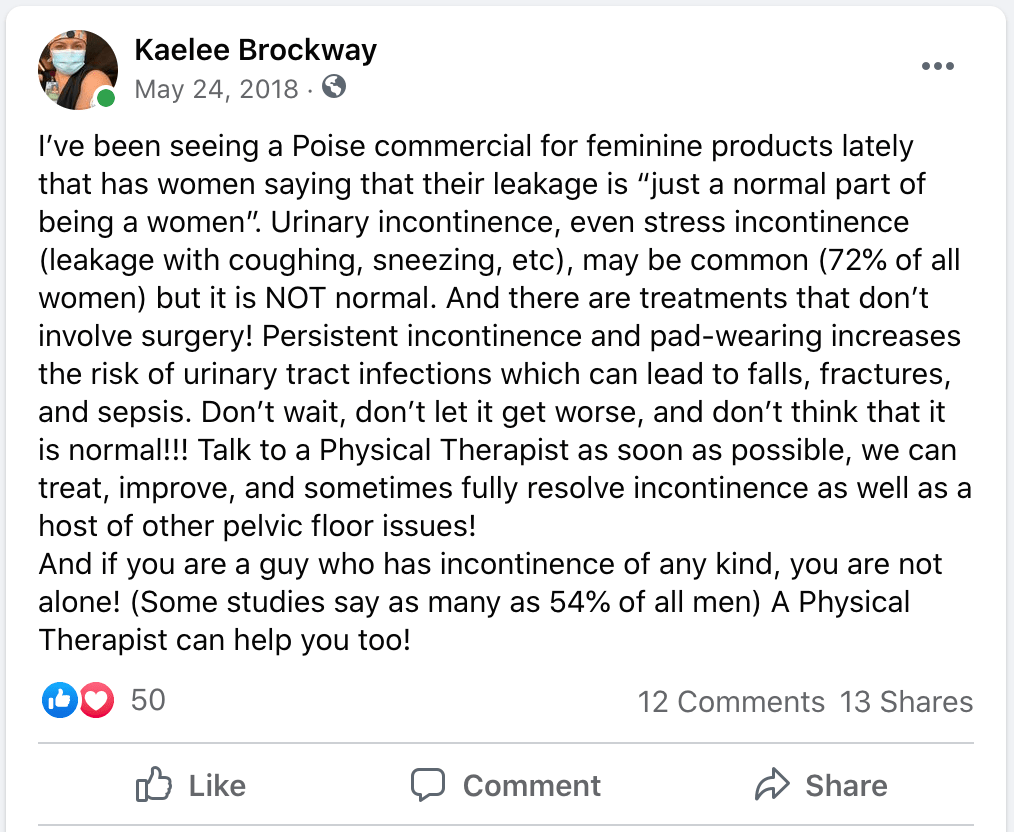
The number one reason I work on intra-abdominal pressure is far and away to reduce urinary incontinence. Think about it. All that pressure inside your abdomen pushing down on… your bladder! The standard method for measuring intra-abdominal pressure is actually to measure it through the bladder! Your bladder can only take so much and eventually won’t stand to the pressure and it will let go. This is called stress urinary incontinence and 80% of women and 45% of men will experience this at some point in their life. In addition to incontinence, this pressure can also cause over-lengthening of the ligaments that hold your organs in place. This can result in herniation of the organs through the abdominal wall (I’m sure you’ve seen plenty of these) or prolapse of the pelvic organs. All things that we ABSOLUTELY DO NOT WANT.

The pelvic floor muscles can normally accommodate to this pressure for short periods of time, but sustained pressure or large amounts of pressure will eventually cause the pelvic floor muscles to either fatigue or lengthen beyond their ability to efficiently contract. After these events, the amount of intra-abdominal pressure that causes incontinence episodes becomes less and less and herniations and prolapses increase more and more. You may be thinking that this is important for post-partum women, but intra-abdominal pressure regulation is also incredibly important for people who have COPD, Heart Failure, Obesity, Cirrhosis, neurologic conditions, and a host of other chronic diseases. That’s because all of these diseases cause changes to intra-abdominal pressure in some way.

Photo by Anna Tarazevich on Pexels.com

For COPD, these patients are coughing frequently. This repeated coughing causes sustained stresses on the pelvic floor muscles and the increased lung volume in obstructive lung diseases increases the pressure within the abdomen. For heart failure, the increased amount of fluid accumulated leads to increased pressure in the abdomen, especially if they experience ascites (accumulation of fluid in the abdomen). Obesity also leads to increased intra-abdominal pressure as the build up of fatty tissue within in the organs takes up space, therefore increasing pressure. With cirrhosis, the liver increases in size due to one of many possible reasons such as edema or fat build up. This takes up extra space in the abdomen, increasing the overall pressure. Tumors or other space-occupying lesions in the abdomen create the same effect. Neurologic conditions can result from increased intra-abdominal pressure, but can also create changes in intra-abdominal pressure. And then, people can have more than one of these conditions and every additional breath can become a turn of the vice. All of these things are becoming so much more common that abdominal hypertension has become a typical diagnosis that can lead to severe health conditions.


Why Do We Need to Regulate Intra-Abdominal Pressure?

Pulmonary conditions are front and center right now thanks to the global COVID-19 pandemic, we will start there. There have actually been cases documented of compression atelectasis (too much fluid in the abdomen increasing pressure so high that the lungs have no where to expand) causing multi-system organ failure. Compression to this level, although somewhat rare, causes internal organ compression to the point of ischemia. This means that all the organ systems can’t get blood because the blood pressure cannot overcome the intra-abdominal internal pressure. As Physical Therapists, we are pretty familiar with compartment syndromes, so to compare, this condition is also known as abdominal compartment syndrome. You can imagine that the intestines, kidneys, and pretty much everything else in the abdominal compartment doesn’t fare well under these conditions. Abdominal compartment syndrome can also be a complication of child delivery.

Returning to our post-partum patients, women who have Cesarean sections (C-sections) need to reduce intra-abdominal pressure when they move to reduce strain on their incision or surgical work. This is also true for most people who have had any kind of abdominal surgery such as hysterectomies, laparatomies, laparascopies, and trachelectomies, and several spinal surgeries such as decompressions, fusions, etc. I’ve also found reducing intra-abdominal pressure to be extremely helpful for people who have had new colostomy placements, as they have decreased pain with movement and decreased complications with their colostomies. We will go in to more detail on all of this later.

Reducing intra-abdominal pressures is crucial in patients with chronic health conditions such as COPD, heart failure, cirrhosis, obesity, and many others. Why, you might ask? They don’t have surgical incisions or internal work that they pressure is pushing on, so what can we do. I’ll take you back to the very first word of this post. You can help them BREATHE. You are essentially providing interventions to improve V/Q matching, because you are allowing increased ability to ventilate and, therefore, increasing potential to diffuse and perfuse. In addition to breathing, most of these patients also experience some kind of incontinence. This is, in large part, due to the increased intra-abdominal pressures, but also due to the effects of medications and altered fluid dynamics. You can’t change the medications or disease, so change the pressure.

Photo by Perchek Industrie on Pexels.com

The neurological population (which we will discuss more in a bit) also struggles with increased intra-abdominal pressures. We will dive deep into the neurologic mechanisms by which this effects the body, but increased intra-abdominal pressure actually impairs the flow of cerebrospinal fluid in the central nervous system. These pressures can also increase venous distension which puts direct back-pressure on the eyes and optic nerve. All of the major neurological components of function are negatively impacted by high intra-abdominal pressure.


Do I Always Want to Reduce the Pressure?

Well, no. Just like everything else in the medical world, nothing is ever “always.” Even lab values that are “not compatible with life” can still be seen in calm living individuals. So, no, you don’t always want to reduce intra-abdominal pressure. In fact, sometimes you want to increase it!

For patients who have medical conditions like Parkinson’s Orthostasis, Postural Orthostatic Hypotensive Syndrome (POTS), or even just your standard orthostatic hypotension, you actually want to INCREASE intra-abdominal pressures, at least temporarily. We achieve this in the short term by actually instructing patients to hold their breath during an activity. This places pressure on their internal vasculature, holding blood up into their brain when gravity says otherwise (like with orthostatic hypotenison: when the patient goes up, their blood – and blood pressure – go down), which can help reduce symptoms of dizziness or blacking out. Obviously we have to let them breathe and return intra-abdominal pressure to normal eventually but we hope to get them active enough to keep the pressure up (usually with some exercise). In the long term, we can use devices like abdominal binders and compression garments to increase the pressure.

For post-partum patients, abdominal binders are also frequently used. This can be for several reasons, but you may actually want to increase their intra-abdominal pressure to apply compression to reduce internal bleeding (there can be a lot of bleeding with and after birth). It is also documented that intra-abdominal pressure can elevate significantly even in healthy pregnancies. This should seem obvious because there is a human growing in a limited space so the pressure has to go somewhere. But, because this is a slow process, pregnant patients tend to accommodate to this pressure over time. However, that tiny human and all it’s stuff leaves the abdomen quickly, which is a harder process to accommodate to. Birth also creates a large fluid deficit which can leave a post-partum patient a bit hemodynamically unsteady. These factors cause the intra-abdominal pressure to drop and some patients may need some pressure support for a while to return to activities normally.

Photo by cottonbro on Pexels.com

Other conditions that cause significant fluctuations in hemodynamic stability, such as severe burns, dehydration, mid-brain injuries, and multi-traumas may also require pressure support for some time.


The Valsalva Maneuver

Some people try to increase their intra-abdominal pressure intentionally on their own. This is typically done using a Valsalva maneuver, or “bearing down”. People use this to place intentional pressure on their organs to achieve a certain goal. This may be to push a baby out, to defecate, or to stabilize their spine. However, using a Valsalva maneuver too often or several times in a row can lead to vasovagal syncope. We mentioned the vagus nerve above and you can see in the drawings below that it provides innervation to the vasculature of the abdomen. Put too much pressure on the vasculature of the abdomen (aka the abdominal aorta) and you will lose pressure support for the blood flow to your brain and you’ll faint. The mechanism also may occur from that pressure causing overstimulation of the sympathetic nervous system resulting in a severe “faint” response, which is your body’s only way of forcing you to stop producing the pressure. It’s basically a safety mechanism to prevent abdominal compartment syndrome.

Alone, vasovagal syncope isn’t dangerous and is usually self-limiting. Just like when you hold you breath long enough, your body will force you to breathe somehow, even if it’s by making you pass out so that you breathe again. However, it’s what happens after you pass out that concerns us. I’ve treated several patients for head injuries, wounds, and broken bones secondary to the fall they experienced during their syncopal episode.

Valsalva Maneuvers are not a great solution to pelvic organ function. This increased pressure to go to alleviate constipation is not effective and has significant side effects. If you are in need of better options, please check out the post on Incontinence Management – Coming Soon!


So What Do I Do To Help These Patients Breathe?

Thankfully, that’s the easy part! Odds are, you already know this, you just may not know the deeper “why” behind why you do it or how to widely apply it. I’m sure you are constantly telling your patients to breathe during their exercises. And I’m sure they repeatedly hold their breath. Mine do! Which tells us they are attempting to stabilize and produce force improperly, i.e. not using their muscles.

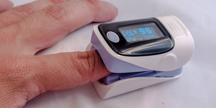
If you read my post on pursed lip breathing, I told you a little bit about this. In that post we talked about how pursed lip breathing, when done correctly, can stimulate the parasympathetic nervous system to reduce anxiety and calm the sympathetic responses. I just used this with a patient recovering from COVID-19 very successfully. When she started to get short of breath and panic, we regulated her breathing and her oxygen saturation sprang right back up. But regulating breathing by changing intra-abdominal pressures can help with so much more! I’m going to take you back to the basics of what you know.

The reason behind this effect is the change in intra-abdominal pressure which changes forces and stimulations to the sympathetic trunks within the abdomen. Applying our anatomy knowledge, we can picture that. Remember the sympathetic trunks from all those years ago? They are coming back. Look at those abdominal sympathetic trunks! They are all over the length of the spinal column from the diaphragm to the pelvic floor and there are more throughout the length of the spine. They feel EVERY change in intra-abdominal pressure and they respond to it. They feel the pressure, they get stimulated and they set off the fight, flight, or faint responses, because that’s their job! Most of the time, we see this manifested as the patient getting anxious, worked up, angry, or refusing to participate. We can also see increased pain or fainting (like we talked about above).

So, by reducing the pressure, thereby reducing the stimulation on the sympathetic nervous system, we calm the fight, flight, or faint response mechanisms. Like we talked about in the post on pursed lip breathing, we also then engage the parasympathetic systems. The parasympathetics mostly branch from Cranial Nerve X: The Vagus Nerve. The remainder of the parasympathetics come from the sacral trunks. Reduce strain on the pelvic floor, and the parasympathetics can do their job.

And, don’t forget, you aren’t just utilizing the autonomic functions to regulate breathing, you are also increasing space for the lungs to expand. This increases the potential for vital capacity, increases the space for residual volume, and reduces the required PEEP, meaning you will have more patent airways. Increased intra-abdominal pressures actually start to require a higher than normal PEEP (positive end-expiratory pressure) to maintain patent airways. Eventually, the body can’t create a high enough natural PEEP to keep breathing properly. Reducing intra-abdominal pressure increases the available space for lung expansion with inhalation, reducing the effort and increasing the efficiency of breathing, and reducing the required PEEP. Significant improvements are noted on Pulmonary Function Tests (PFTs) including improved FEV1 and improved FRC, both markers of overall pulmonary function, when intra-abdominal pressure is reduced.


How Can I Reduce the Pressure So These Patients Can Breathe and Move Better?

After spine or abdominal surgery or birth, patients are NOT taught how to move! They strain. They hold their breath. They endure. Or they don’t move at all. Patients who have chronic diseases have been slowly accommodating to increased intra-abdominal pressure and changes in lung volumes and compliance over time, so may not feel an acute change in their symptoms. But, all of this is 100% avoidable. And, oh so simple. This is where you come in.

If your patient is having pain with movement post-operatively, go to your basic techniques and then we will build on them a bit. We know for post-operative spine patients, log rolling is very important to prevent them from breaking their movement and positioning precautions, but this concept can be applied broadly. Especially for patients who have abdominal incisions, we need to decrease the tissue strain or they are going to hold their breath because of pain. Then, we have to pair breathing with the movement to reduce sympathetic activation and reduce intra-abdominal pressure so that muscles can function properly with an appropriate vitals response. Here are the steps for something simple:

Getting out of bed
  1. Start in Supine (lowest intra-abdominal pressure)
  2. As they breathe out: Slide one foot up the bed while breathing out through the whole motion, breathe in
  3. As they breathe out: Slide the other foot up the bed while breathing out through the whole motion (this brings you to hooklying), breathe in
  4. As they breathe out: As a whole unit, roll on to one side while breathing out through the whole motion, breathe in
    1. Patient may need to use grab bars, trapeze, bed rails, drop knees to the side, UE support from you, or momentum from swinging UEs
  5. From sidelying, position hands near head and shoulder on bed to push up to sitting and bring feet forward over edge of bed while breathing out
  6. With or without assistance, push up from the arms and drop feet downward to the floor while breathing out through the whole motion (this achieves sitting which created only small increases in intra-abdominal pressure in comparison to supine).

I know that seems like a lot of steps, but it moves rather fluidly once you teach it. It is so important to break the task down and ensure the patient moves slowly and breathes out (exhales) with each position change. This reduces the intra-abdominal pressure during movement, keeps tissue strain to a minimum, and also keeps that sympathetic nervous system quiet so the patient can focus on moving. If your patient uses supplemental oxygen, make sure they are breathing in through their nose so they can utilize it most effectively to maintain V/Q matching. And, even though this is a lot of steps which increases the time required to perform this activity, your patient will thank you for the increase in their comfort. They won’t have to do it this way forever, just until they recover from their surgery.

Now you can take these concepts and apply them widely, like with sitting down on the toilet, or getting up from the floor, or with performing their exercises in the gym like their next plyo task or weighted deadlift. If your patient experiences pain or difficulty breathing with movement, you’ll likely see a decrease in either or both symptoms because of those mechanisms we discussed above. In the post-partum population, you may find that your patient needs both pressure support and pressure reduction depending on the movement task.

If you are using intra-abdominal pressure regulation to help treat your patients with stress incontinence, there are some specific ways you need to build breathing in to you exercises and other treatments. Remember how we talked about the pressure this places on the pelvic floor? Yea, you have to reduce that pressure if you want your patient to have the proper length-tension relationships in their pelvic floor muscles to even attempt a proper muscle contraction. It is so often that people hold their breath while performing pelvic floor exercises. I like to tell my patients that those particular muscles are NOT breathing muscles. They laugh, which then means they are breathing, which makes me happy, and then we try again. I think that leads me to my next post on incontinence management… See you there!


Even if you already knew about these interventions, I’m hoping that you now can better utilize them for a wider variety of patients. I also hope you have a really good understanding of WHY you use these techniques. How do you use intra-abdominal pressure regulation in your patients? Tell me in the comments!

Follow my blog for more!

More Reads…

Dehydration

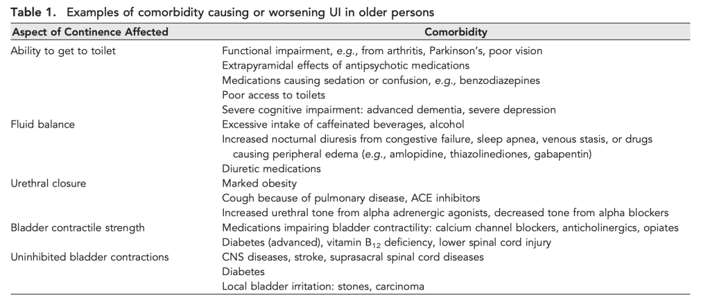
WHILE WE WAIT FOR THE NECT CHAPTER OF DIABETES MANAGEMENT, LET’S KEEP TALKING ABOUT INCONTINENCE Chronic management of urinary incontinence can lead to many issues like infection and hospitalization if it doesn’t account for fluid balance! Let’s talk I’s and O’s! #physicaltherapy #incontinence #chronicdisease

Cobb, W.S., Burns, J.M., Kercher, K.W., Matthews, B.D., Norton, H.J., Heniford, B.T. (2005)
Normal Intraabdominal Pressure in Healthy Adults. Journal of Surgical Research. 129(2):231-235. https://doi.org/10.1016/j.jss.2005.06.015.

Depauw, Paul & Groen, Rob & Van Loon, Johannes & Peul, Wilco & Malbrain, Manu & De Waele, Jan. (2019). The significance of intra-abdominal pressure in neurosurgery and neurological diseases: a narrative review and a conceptual proposal. Acta Neurochirurgica. 161. 1-10. 10.1007/s00701-019-03868-7.

Frezza, E.E., Shebani, K.O., Robertson, J. et al. Morbid Obesity Causes Chronic Increase of Intraabdominal Pressure. Dig Dis Sci 52, 1038–1041 (2007). https://doi.org/10.1007/s10620-006-9203-4

Mayo Clinic. (2021). Vasovagal Syncope. Retrieved from https://www.mayoclinic.org/diseases-conditions/vasovagal-syncope/symptoms-causes/syc-20350527#:~:text=Vasovagal%20syncope%20(vay%2Dzoh%2D,blood%20pressure%20to%20drop%20suddenly.

Sugerman, H.J., Bloomfield, G.L. & Saggi, B.W. Multisystem organ failure secondary to increased intraabdominal pressure. Infection 27, 61–66 (1999). https://doi.org/10.1007/BF02565176

Follow @DoctorBthePT on Twitter for regular updates!

Dehydration

Gosh, this is a fine line… Especially in the patients I regularly see. A colleague and I often say, “If you have any more water, you’ll die. If you don’t have any more water, you’ll die.” This is actually a frequent education topic that I cover with patients. Scary? Yes, but true. So, why is a Physical Therapist going to talk to you about dehydration and water intake? I cannot even begin to explain to you in any short way how important this is to my DAILY practice.

Symptoms of dehydration include:

  • dizziness
  • light headedness
  • low blood pressure
  • orthostatic hypotension
  • increased thirst
  • fatigue
  • confusion
  • falls
  • urinary tract infections
  • incontinence
  • lack of sweating (in severe cases)
  • loss of consciousness (in severe cases)
  • cardiac arrhythmias (in severe or chronic cases)
  • loss of thirst (in severe cases)
  • the list goes on…

I treat all of those things as symptoms of other conditions and some in isolation. I bet you do, too!

Acute, chronic, or severe dehydration results in hypovolemia, which means that your body doesn’t have enough volume (blood volume) to support normal function. Guess what, folks? Blood pressure is a function of blood volume. So, if you lose blood volume, you will also lose blood pressure. When you lose blood pressure, your heart rate has to increase to make up for the loss in cardiac output to keep you going. This results in vitals findings that include a low blood pressure and an elevated heart rate. This is part of the discussion around hemodynamic stability, one of my favorite things to talk about.

For example, I’ve had a patient just recently that demonstrated blood pressures at rest 96/54 mmHg and a heart rate of 104 bpm. Sometimes, you don’t see the change in blood pressure and heart rate at rest, but you may see it with activity. The other day, I was teaching an exercise test and our model had resting blood pressure of 120/86 mmHg and a heart rate of 70 bpm. With ONE MINUTE of marching in place, her blood pressure was 130/90 and her heart rate was 50 bpm. She was also having cardiac arrhythmias that were not present at rest. It took ONE MINUTE. That’s it. And, just an FYI, this was NOT a patient. This was your standard run of the mill adult walking around doing their normal thing. This adult could be walking in to your clinic at any moment.


Why, you might ask, are so many of Doctor B’s patients dehydrated? Well, there are lots of reasons. I’m going to use this post to segue into a few other topics, but the gist of it is that they are sick. And when you are as sick as they are, drinking enough water is either difficult or not indicated. That’s right. You read that correctly. For some of my patients, drinking too much water is actually exactly their problem. You can read more about that here. But the REAL REASON that many of my patients are dehydrated is that I check for it. Simply because I check for it, I’ve significantly increased the number of dehydrated adults on my caseload. Just because someone is sick doesn’t mean they are dehydrated, but if you never check, you’ll never know.

Many of my patients have difficulty breathing. Many of them have COPD or some other form of lung disease. If it’s difficult to breathe at rest, getting up to get a drink of water is going to be pretty challenging, so many of them just don’t. For people who use supplemental oxygen, staying hydrated is even more important because their supplemental oxygen is very dry, even when using a humidifier, and causes cracks and bleeding in their nose. This causes discomfort and decreases compliance with their oxygen.

Photo by Daria Shevtsova on Pexels.com

If you’ve read my posts on heart failure, including the post that covers the Rule of 2s in relationship to heart failure, you can understand how too much water can actually become a problem. For patients who manage chronic heart failure, their fluid restriction (typically of 2 liters of water per day) can make them fairly dry, especially in the beginning of their disease. They are used to drinking as much water as they need, but all of a sudden, we are telling them they have to ration it out over the course of the day. Initially, they have to adjust to this change so they have some time frame of mild orthostasis or other dehydration symptoms. Funnily enough, they usually do fine for a while, but then they get too good at limiting their fluid intake and they start to REALLY limit themselves. Some patients are proud to announce that they can only drink 1 liter of water per day… And Doctor B cringes… Even when they have a fluid restriction, it is still important to drink to the max of their restriction to ensure they stay hydrated enough to function.

Many of my patients are also incontinent. We will talk more about this later, but the solution many of them think they’ve discovered is that, if they drink less water, they won’t have to get up to use the bathroom as often (which is hard for them to do, or requires the assistance of another person and they don’t want to be a bother). Unfortunately, this results in these older, more fragile people becoming dehydrated which produces hypovolemia, which results in increased sodium and potassium concentrations in their body (hypernatremia/hyperkalemia), which results in altered mental status and/or heart problems. Unfortunately for them, drinking less water doesn’t actually reduce their urination frequency….

That’s right, decreased water intake doesn’t really decrease your urinary frequency. What actually happens is that your urine becomes more concentrated in your bladder. This concentrated urine becomes a bladder irritant which actually increases the frequency of feeling the need to go! This is especially true of those middle-of-the-night trips to the bathroom that we all know result in falls. People stop drinking water an hour or two before bedtime so they won’t have to get up to use the bathroom at night. But, darn it, that’s just not how it works. Now they will actually be getting up more often to use the bathroom because they are getting dehydrated before bed.

Photo by Pixabay on Pexels.com

Let’s factor in how often urinary incontinence co-occurs with chronic diseases, especially those of the respiratory system. You don’t have to go out and do your own research, I’ll just tell you: It’s pretty much every time. My patients often have to cough to clear secretions, because their throat is too dry, or because they have swallowing impairments. When they cough they become incontinent. They have trouble getting up so they just don’t. Then, they sit in that extra-concentrated urine until they muster the energy or someone is available to help them. They end up developing urinary tract infections which take them down the long spiraling road of confusion, falls, injuries, and hospitalizations.


Setting doesn’t matter. Many of my chronic disease folks are in the home health setting, but I see plenty of dehydration in outpatient orthopedics, too. Even in the Skilled Nursing Facility where the nurses do daily rounds to check vitals, many more than once per day, I still check every patient before exercising them. Patients in facilities tend to be pretty dry, partially because they can’t get more to drink on their own. Finding patients too orthostatic to participate in therapy and recommending fluids has become a regular intervention.

I think you can all pretty much get the picture as to why not drinking enough water is really bad, especially for the older population or those with chronic diseases. And the threshold is pretty narrow for many people if they also have a fluid restriction. But, do you remember why I said so many of my patients have dehydration? The REAL reason? It’s because I check for it. And so should you.

Vitals. Every patient. Every time. More than once.

And you can be the one to prevent that hospitalization or that infection or that fall. All you have to do is look for it to know it’s there. Then you can intervene and be the hero with just a cup of water.


How many cups of water do you drink every day? Let me know in the comments!

Pressure… Pushing Down On Me…

Breathing. I can’t stress it enough. If you’re not breathing, you’re dead… or in a lot of pain… either way, it’s not good. So breathe! In my practice, I work with a lot of different types of patients with a wide variety of conditions and comorbidities, but they all have one thing in common: they…

Home (Health) is Where the Heart Is

Dr. Rebekah Griffith, other wise known as The ED PT, interviewed Dr. B on all things patient care management and how PTs in the ED and Home Health PTs can work together to provide the best possible care for patients. We talked about the struggles we each face in managing complex patients and the roles…

Loading…

Something went wrong. Please refresh the page and/or try again.

Follow @DoctorBthePT on Twitter for regular updates!

Chronic Disease Part 3: Urinary Incontinence – Part 1

All that work on intra-abdominal pressure really kept taking me down the road of incontinence management, but that post was already long enough, so this one is going to get it’s own feature. I work with patients on managing urinary incontinence in every setting, so let me take you through some basics of how to do this. I am not a certified pelvic floor Physical Therapist and I only claim to know the absolute basics, but for many people, the basics are pretty helpful, and can at least buy time until they can get to a specialist if they need to. You may be thinking, “Why is this person who does mostly cardiopulmonary all day want to talk about urinary incontinence?” Well, most of my patients who have cardiopulmonary conditions like COPD or Heart Failure either have secondary effects of those conditions that cause incontinence or take medications that cause incontinence. So, out of necessity, I had to learn how to address these things.

This is a huge topic, also, so it’s going to be broken in to two parts. The first part is heavy on the what and the why so that you can have a really good understanding of the importance of managing this condition in patients across different setting. This will also help you have some great ideas for how to document what you are doing and why.

Why Is Urinary Incontinence Such a Problem?

Urinary incontinence straight up leads to disability. It may not be immediate, or it may be, but it’s a slippery slope and a short road. Urinary incontinence is extremely common, but it is NOT normal. It happens to both men and women, but definitely more often in women. When I was in PT school, we learned that about 80% of women will experience incontinence of some kind at some point in their life. Further research showed that this is also true of about 54% of men. That’s because incontinence doesn’t just come from childbirth, believe it or not. You can read more about things that cause stress incontinence in the post about intra-abdominal pressure, but we will go in to some detail here.

Urinary incontinence is the #1 reason women are placed in a facility as they age. That is because it becomes pretty hard to manage. There are many products that require purchasing, so it also becomes expensive. Incontinence is also among the leading causes for institutionalization of men as they age. This facility placement also typically leads to these men and women becoming dependent in their toileting, because they transition from what was otherwise a stress, urge, or mixed incontinence, to a functional incontinence. You can read more about that below, but basically, if there is no one available to help you, you eventually get to a point where you just go. This has been shown to increase depressive symptoms and severely impact overall well-being. Urinary management was the most-often reported impaired ADL in people over 65 years old, but it happens at ANY age.

And, finally, it is all potentially preventable. All of the journals cited in this article and several geriatric organizations agree that promoting continence or management of continence in the geriatric population is the most important factor in maintaining independence and well-being. But, it’s not just about the geriatric population, right? I did plenty of treatment for incontinence in outpatient orthopedics for patients of all ages, both men and women. Unfortunately, we often don’t see younger patients in therapy specifically for incontinence, but they do tend to come in for other issues… specifically back pain, hip pain, and core weakness! The incidence of stress urinary incontinence coexisting with low back pain is high, especially in women. This is where you come in. Let’s learn a bit more about Urinary Incontinence.

Types of Urinary Incontinence
  • Stress. We talked A TON about this stress in the article on intra-abdominal pressure regulation, so you can the details there, but overall, if you place enough stress on the bladder, it will empty. This type of incontinence happens when you cough, sneeze, stand up, hold your breath, turn over, strain… pretty much anything that can increase intra-abdominal pressure. This type is more common in women than in men, but, if you’ve seen men with abdominal hernias, you know it exists in men, too.

  • Urge. Sometimes called “key-in-door” incontinence, this type comes from a sudden and unexpected urge to urinate. This urge may be neurologic or reflexive in nature, like you always have to go when you get home from work so when you get to the door, you have the feeling that you have to go. It has been shown to be due to the combination of some type of reflexive or neurogenic trigger AND an overactive detrusor muscle (the muscle that surrounds the bladder). This urge can sometimes come from bladder irritation, too, and we will talk about what that means later. This type is actually more common in men than in women.

  • Mixed. This is the descriptor for some type of combination effect of both stress and urge incontinence.

  • Overflow. This one is pretty self-explanatory. When your bladder gets too full, it HAS to empty, whether you are ready or not. Sometimes, you just wait too long. Or, as with otherwise potty-trained children, they can get distracted by playing, and then they just go. Overflow incontinence can also happen when bladder emptying is impaired. This can happen by many mechanisms such as prostate enlargement, scarring of the urethra, detrusor weakness, or a bladder prolapse (cystocele). If the exit is blocked, nothing comes out.

  • Functional. This is the worst type of incontinence because it is 100% preventable. Functional incontinence is incontinence that happens when you can’t get to a bathroom safely or don’t have the help to manage the bathroom tasks that you need to perform. It can also result from shame, embarrassment, weakness, balance impairments, homelessness, and environmental barriers. Here is a clip of me talking about functional incontinence so you can get an idea of how this happens:

    Doctor B talking about functional incontinence in the aging population
  • Neurogenic. Neurogenic bladder results from some type of neurologic disruption to the bladder and its systems. This could be in a patient with spinal cord injury who cannot feel that their bladder is full. They no longer have the connections to the reflex mechanisms that send their brain that signal, so they have to perform intermittent catheterizations to empty on a regular schedule. Other neurological conditions, like Multiple Sclerosis, Cerebrovascular Accidents, Parkinson’s Disease, and many others can impact these reflex loops or their centers in the brain and result in a neurogenic bladder. Trauma can also lead to this condition if there is damage to the sympathetic (T10-L2) or parasympathetic (S2-4) trunks that innervate the detrusor muscle.


And, as with many things, the likelihood of urinary incontinence increases with age. This is due to many factors, but the compounding of these factors, like the increased number of comobordities accumulated with age, increases the risk of urinary incontinence. There are several health conditions that result in different types of urinary incontinence by direct of indirect mechanisms. The American Society of Nephrology produced this great table that gives you a good overview of the things that can cause urinary incontinence.

American Society of Nephrology

I want to point out, specifically, the line in this table on “Bladder contractile strength”. There are so many things that change the ability of the bladder to contract appropriately, and most of my patients have at least one of the items on that list in their medications list of past medical history. The bladder is surrounded by a muscle, right? And it has neurological inputs and outputs and can be strengthened, just like any other muscle! And you are a Physical Therapist, isn’t that what you do? So let’s take the first step:

Identifying Urinary Incontinence

No one really wants to talk about it. No one really wants to admit it. So, you can’t just ask, “Do you pee your pants sometimes?” because it won’t get you any useful information. In the home setting, it’s probably most beneficial to just look around. You’ll see what you need to see if someone has incontinence. There will be products readily available in the bathroom, and there may even be soiled clothing laying about. In the outpatient setting, it’s a bit tougher because you can’t take as many nonverbal clues. You have to get crafy in your questions! Here’s how I go about this:

“Do you ever have any urine leakage?”
“Do you ever have any other type of leakage?”
“What about when you cough or sneeze sometimes?”
“Do you ever need to use any products for leakage like pads or undergarments?”
“How often do you need to change those in a day?”
“Do you notice you have leakage with any certain activities?”


And just because they answer “No.” to the first one, doesn’t mean I don’t ask the others. Often times I will get “No,” as the first answer, but then yes to several of the other questions. It is also incredibly important to follow up this series of questions with a conversation about how, although this is common, it is NOT normal, and can be improved or resolved very easily, even if it has been happening for a long time. You may also need to add a conversation about how pads and undergarments are only a band aid, not a solution, but we will get more into that later.


How is Urinary Incontinence Treated?

Well… here’s the thing. Most people who have it don’t treat it. They simply think it’s normal. Most of them are women who have had children and they just think that’s how things are. BUT IT’S NOT TRUE. Other treatments tend to focus on type and cause. Normalize the situation, but also normalize treatment. Many people will be ashamed or embarrassed and they will need you to coach them through that in order to make progress toward a solution.

Using liners, pads, or briefs allows urine and other body waste to stay in contact with the urethra, resulting in some of it working it’s way back in. The result, especially in older adults and those who are immunocompromised, is a urinary tract infection. Urinary tract infections (UTIs) are incredibly common (but also NOT normal) and the incidence is rising. This is a much bigger problem than most people realize and here’s why: Antimicrobial resistance, also known as super bugs. The number of hospitalizations for UTIs is rising year after year, especially over the summer months, and doctors think that this is due to patients being previously treated for UTIs but returning due to a developed resistance to antibiotics.

Urinary tract infections are hugely problematic. They are incredibly dangerous and can result in sepsis, falls, temporary or permanent mental status changes, and continually decreasing urinary health and function. All of which results in decreased functional ability, hospitalization, institutionalization, or even death. They also contribute to the aforementioned building concern for drug-resistant microbes. I may not be a pelvic floor therapist, but I’m pretty darn vocal about this! Here are the types of urinary incontinence and how they are typically treated at the present. Hopefully we can make some serious changes!

Stress incontinence. As a secondary effect of other comorbidities, stress incontinence can be treated with medications and other interventions for the comorbidities, along with retraining of how they regulate their intra-abdominal pressure. As a primary diagnosis, regulation of intra-abdominal pressure and pelvic floor strengthening exercises are usually the keys to success.

Overflow incontinence.
Treated by removing whatever blockage is present to the urethra. This typically involves surgical repair for prolapses, bladder suspension, or prostate removal. Unfortunately, these procedures come with a laundry lists of side effects and effectiveness is questioned. If the blockage cannot be removed, intermittent catheterization or indwelling catheters can be used, but have serious potential for problems.

Urge incontinence. May be due only in part to an overactive detrusor muscle, but is typically only treated using medications that target relaxation of the detrusor muscle. Unfortunately, this can result in over-relaxation of all the muscles or just the detrusor muscle which then results in several other issues. And, this treatment only treats half the problem! We now that there is a neurological component to urge incontinence.

No Treatment. Most commonly, there are those who don’t treat it, but attempt to manage it using liners, pads, or briefs, effectively allowing it to happen, but not letting anyone else know it is happening. In men, catheterizing is a common technique to address incontinence. Here’s the problem with all of that:

Infection.


This is Serious! I Need to Treat This! What Do I Need to Do?

Physical Therapists play a very large role in managing urinary incontinence. If you read through that table from the American Academy of Nephrology, I’m sure you noticed that most things on the list are things we can address in some way, whether it is a medication reconciliation, a lifestyle change, and environmental change, or physical intervention. There are going to be LOTS of options for you to chose from for your patients. There is something for everyone! So make sure to check back!

Reduce modifiable factors

Some things you can change, some things you can’t. So work with what you can change. You can’t change someone’s age, gender, or disease. You can change their location (maybe), their available help (maybe), their medications (maybe), their mental state and function (maybe), their BMI (maybe), their safety, their functional ability, their strength, their diet, and their environment. A lot of maybes… But this gives you many different avenues from which you can launch your treatment. Let’s attack the maybes first:

If you band together with some of your healthcare team, you can address some of these maybes pretty well. You can work with a social worker to get in-home help or find suitable supportive placement. I know that institutionalization is not the best answer, but sometimes it’s the only answer, and sometimes its better than being alone and unsafe, but you can also consider short term rehab facility placement. You can work with the primary care doctor (or urologist and cardiologist, if you are lucky) to change medications or reduce dosages to reduce cognitive effects, stabilize fluid dynamics, and reduce fall risk. You can work with family to arrange for in-home assistance. And, as a skilled rehab provider, you can get someone going on an exercise program to begin chipping away at their BMI.

Other than the maybes, we have some pretty tangible stuff in this list that Physical Therapists are pretty poised to address. These are some of the things I was talking about when I mentioned the lifestyle changes. Let’s take a look at those:

01


Environment

Take a look around. Reduce barriers, ensure safety. Turn lights on, make paths short and straight. Make sure they have the appropriate assistive device.

02


Functional Ability

The path is great, but we have to get to it. Make sure they can get out of a chair by elevating it if needed.

03


Diet & Fluids

More water is better. I know this is counterintuitive, but it’s important. Reduce or eliminate bladder irritants.

04


Strength

Lower extremity strength, Upper extremity strength, core strength, pelvic floor muscle strength… Pick one or several, depending on your patient’s needs.

Bladder Irritants? Yes, that’s a thing.

Over-concentrated urine is a bladder irritant, so cutting water intake should not be an option. Keeping a patient with heart failure or ESRD within their approved water intake is important, but reducing it is not a solution. Many patients like to cut out liquids before bed so they won’t have to get up to use the bathroom at night. Although this strategy sounds plausible, it results in increased urine concentration and your patient will actually have to get up more often. Patients who are returning to a healthy water intake after restrictions will initially experience an increase in urinary frequency, but their body will regulate with time. If your patient doesn’t like water, have them gently flavor it with flavor drops (not powder or sugar-based drink flavors) or fruit.

Some food and drinks are also bladder irritants. As much as it pains me to say it: Coffee. Technically, caffeine is the culprit, though. Let me tell you this little pearl: DO NOT tell your patient to stop drinking coffee. You’ll lose them immediately. Ask them, instead, to alternate a cup of coffee with a cup of water. Other highly caffeinated drinks like soda are also on this list, as well as highly acidic foods and liquids. Citrus fruits, tomato products, and spicy foods will all result in increased urge or frequency or both. However, diluting these irritants can help, so keep the water flowing.

Another one that will get you gaffs if you suggest it’s removal is alcohol. Alcohol, much like coffee, dehydrates you, increases urine concentration and causes bladder irritation. Like I said with coffee, don’t suggest eliminating it, suggest diluting it by alternating with water. If your patient is treating their urinary incontinence with medications, you’ll want to advise removing alcohol completely as it has severe interactions with some of these medications.

Bladder Retraining Programs

This is where a referral to our magical friends in the pelvic floor rehab arena are called upon. We create voiding schedules for our patients with spinal cord injury, and this process is similar. If you have established a reflexive (literally, neurological reflex loop) habit of going to the bathroom at a given time of day or before/after a certain activity, your brain will learn this pattern and make it happen. These schedules can be helpful to maintain continence, but they can also be detrimental if they are not created intentionally. Remember that “key-in-door” incontinence we talked about? What if you can’t find your keys? Problem! Try going before you leave work. Then you’ll know you don’t actually have to go and you can calm your jets.

It is extremely important to remember that our body has an alarm system for a reason. Our bladder sends signals to our brain to say, “Hi there, I’m full! Time to empty me. Better find a bathroom soon.” If we don’t respond to these signals, they will intensify, “Hey, I’m telling you I gotta go, so let’s go! Now!” And as long as you get there, that’s great. The system worked. But this can go awry in two ways.

  1. What if you can’t get there?
    This is where functional incontinence happens. If you can’t get there even though you know you have to go, you need to identify the reason. Strength, environment, endurance, etc… See the list above. If you don’t get there, overflow happens and you go anyway.
  2. You keep ignoring the signals.
    This actually creates a negative learning loop. If you ignore the signals enough, the body and brain figure out that the signals aren’t working and the bladder stops sending them. BAD PLAN. Now how the heck are you going to know when you have to go? And we end up in overflow again with no warning.

Your local pelvic floor therapist is going to be the best one to help you figure out these schedules, reflexes, and learning loops to best set up a schedule that fits your patient’s personal needs. Don’t forget that incontinence happens at all ages, so we may need to consider school and work schedules when timing bathroom breaks.

Finally… Pelvic Floor Muscle Strengthening and other Bladder Management Techniques

This is where I’m going to leave you in a bit of suspense! This post is already getting pretty long and complex, so I’m going to make treatment options an entirely different post. Stay tuned! I’ll link Part 2 once it is published.


So much to think about and so many people we can help! I once was able to resolve functional incontinence in a patient simply by having her turn on the bathroom light prior to entering. It can be that simple! Do you have a story about a lifestyle change that significantly improved incontinence? Tell me your story in the comments!

Follow my blog for more!

More on How Physical Therapists Manage Chronic Disease!

Treating Long-COVID: Part 2

NEW POST COMING MONDAY! But I can’t give you Part 1 of Treating Long COVID without giving you Part 2! #covid #physicaltherapy #chronicdisease

Treating Long-COVID: Part 1

After spending the weekend learning about the connections between COVID, Long COVID, and other health conditions, we just have to talk again about COVID and diabetes! Check it out!

That Beautiful Teaching Moment: ABI

NEW BLOG POST COMING SOON!! We will continue on our journey through diabetes management! And what comes along with Diabetes? PVD of course! Let’s talk about managing some of the side effects of diabetes…

Loading…

Something went wrong. Please refresh the page and/or try again.

References

Ayeleke, R. O., Hay-Smith, E. J., & Omar, M. I. (2015). Pelvic floor muscle training added to another active treatment versus the same active treatment alone for urinary incontinence in women. The Cochrane database of systematic reviews2015(11), CD010551. https://doi.org/10.1002/14651858.CD010551.pub3

Bush, H. M., Pagorek, S., Kuperstein, J., Guo, J., Ballert, K. N., & Crofford, L. J. (2013). The Association of Chronic Back Pain and Stress Urinary Incontinence: A Cross-Sectional Study. Journal of women’s health physical therapy37(1), 11–18. https://doi.org/10.1097/JWH.0b013e31828c1ab3

Carvalho, N., Fustinoni, S., Abolhassani, N. et al. Impact of urine and mixed incontinence on long-term care preference: a vignette-survey study of community-dwelling older adults. BMC Geriatr 20, 69 (2020). https://doi.org/10.1186/s12877-020-1439-x

Kuchel, G. A. and DuBeau, C. E. (2009). Urinary Incontinence in the Elderly in Geriatric Nephrology. American Society of Nephrology. Retrieved from https://www.asn-online.org/education/distancelearning/curricula/geriatrics/Chapter30.pdf

Mayo Clinic. (2020). Bladder Control: Lifestyle strategies ease problems. Retrieved from https://www.mayoclinic.org/diseases-conditions/urinary-incontinence/in-depth/bladder-control-problem/art-20046597

Nuotio M, Tammela TL, Luukkaala T, Jylhä M. Predictors of institutionalization in an older population during a 13-year period: the effect of urge incontinence. J Gerontol A Biol Sci Med Sci. 2003 Aug;58(8):756-62. doi: 10.1093/gerona/58.8.m756. PMID: 12902536.

Simmering, J. E., Tang, F., Cavanaugh, J. E., Polgreen, L. A., & Polgreen, P. M. (2017). The Increase in Hospitalizations for Urinary Tract Infections and the Associated Costs in the United States, 1998-2011. Open forum infectious diseases4(1), ofw281. https://doi.org/10.1093/ofid/ofw281

Welk B, Baverstock R. Is there a link between back pain and urinary symptoms? Neurourol Urodyn. 2020 Feb;39(2):523-532. doi: 10.1002/nau.24269. Epub 2020 Jan 3. PMID: 31899561.

Zilberberg MD, Shorr AF. Secular trends in gram-negative resistance among urinary tract infection hospitalizations in the United States, 2000–2009. Infect Control Hosp Epidemiol 2013; 34: 940–6.

Follow @DoctorBthePT on Twitter for regular updates!

Chronic Disease Part 4: Type 2 Diabetes – Part 2

Make sure to check out Part 1 of this series HERE!

Let’s Jump Right In

And… moving on! I made some promises in the last post about the diabetic foot, diabetic ketoacidosis, insulin sliding scales and other medications, and using exercise to treat diabetes, so we are going to go through a few posts to cover those topics and get you ready to fully manage folks who have Type 2. This part of the series will focus on diabetic foot management.

As PTs, we frequently look at, touch, and work with people’s feet. Most people have feet and our feet are the base of our body. There is an older book out there called When the Foot Hits the Ground, Everything Changes and it was a standby resource at one of my old clinics. Needless to say, the feet are pretty important. But some populations, the diabetic population, in particular, have a pretty high risk of losing their feet (or parts of them). Amputees are a large part of my caseload and I can say that with certainty across any setting. Home health, skilled nursing, subacute rehab, inpatient rehab, acute care, outpatient… yep, lots of missing parts. Not all of them are due to diabetes, but in the Geriatric population, many are. And those are my people.

So, let’s talk about feet!

Photo by Pixabay on Pexels.com

The Diabetic Foot

Because the feet are at the bottom of the body, they are typically the furthest from the heart (or the pump) and, therefore, receive blood with the assistance of gravity (because gravity helps the blood go downward). As blood vessels get further from the heart, they also tend to get smaller, and if you do some basic math, that means that the blood in your feet will likely be under higher pressure than the blood in your arms, but the difference shouldn’t be too far off in a well-functioning system. You can read more about that here.

Now, those smaller blood vessels have a lot of work to do, supplying all the small muscles, nerves, bones, and other tissues in the feet, and then return all the waste products from those tissues back to the removals systems in our body. Getting blood down to them is not usually an issue, but because blood has to work against gravity to return to the source (the heart), our body has valves in the veins to help keep the blood from flowing back down to the feet after it has left. This process is also assisted by muscles in the legs that squeeze the arteries and veins as they contract and relax. If you’ve ever been told about ankle pumps or have taught someone to do ankle pumps, this was probably the reason behind that activity: help pump blood, waste, and fluid out of the feet and against gravity.

So far, this should all sound pretty basic, which is great! because most of these things to this point aren’t that complicated. However, when you add diabetes to the mix, things start to change and the whole system begins to become far more complicated. Let’s talk about peripheral vascular disease for a minute.

Photo by EVG Kowalievska on Pexels.com

Now, I could talk about vascular disease pretty much all day, but the gist of it is that when blood glucose (or “blood sugar”) levels go unchecked, this causes damage to the endothelium (the most intimate and fragile layer of the blood vessels). This damage results in scar tissue and other things that shouldn’t be there getting deposited within the wall of the blood vessel. This scarring and buildup cause the blood vessel walls to harden and, eventually, the muscles within the walls of the arteries do not work anymore. The artery just becomes a stiff pipe that cannot respond to increased or decreased demand for blood and oxygen from the tissue below (in the foot). The tissues of the foot are then starved for these vital substances and cannot maintain good tissue health. And now we’ve gotten to where we are going… It’s a bit of a long road and it takes some winding turns, but let’s talk about what all that means. We will go by tissue type:

Nerves

Nerves require a blood supply to function. Nerves are also myelinated (or covered in fat) which helps them carry signals faster than they otherwise could. This fat also requires blood supply. When nerves don’t get the oxygen and nutrients they need, they can’t send signals properly or quickly, which can result in erroneous signals or a lack of signals. In patients with diabetes, we often hear this called neuropathy. And, in people with diabetes, this neuropathy happens distal (or furthest away) to proximal (closest) resulting in a stocking-style presentation of paresthesias or anesthesia.
Of note, in the upper extremities, it can present in a glove-live pattern. I see this more often in people who have Type 1 diabetes, but we are talking about the feet here.

If the nerves aren’t sending signals properly, or at all, the foot can’t feel the sock or shoe it is in. This can result in pressure sores from improperly fitting shoes or broken-down shoes. Wounds can get infected pretty easily when they are on the feet, so sepsis ensues and this is how feet are lost. The foot also can’t feel the ground, and this is a big one because that implies effects all the way up to the brain. Here’s what I mean: If the foot can’t feel the ground, then the brain has to use other mechanisms to ensure it is on the ground, like the vestibular and visual systems. These three normally all work together, but now only two are working properly, so that patient has lost one-third of their balance systems. Now we have an increased risk of falls.

Photo by Thought Catalog on Pexels.com

Just as there are blood vessels to nerves, there are also nerves to blood vessels, so their function is intricately intertwined. There are intimate interactions between the nervous and vascular systems, and malfunctions in one will result in malfunctions in the other. This also contributes to neuropathy and several other neurovascular issues throughout the sympathetic and parasympathetic nervous systems which impair the body’s internal management of insulin and blood glucose. But that’s a complex topic for another time.

Skin

If skin cannot get a good supply of blood, it will have to retreat its resources to the deeper layers that protect organs and sacrifice the superficial layers. This results in patients with diabetes having very dry, fragile, and flakey skin, especially on their feet. It is a common part of the diabetic foot assessment to remove a sock and find a snowbank worth of skin flaking off. I know, it sounds gross, but the body just can’t supply all the layers, so they shed off faster. This also means that the hair follicles die and sweat glands are dysfunctional. The skin also breaks down much faster due to the increased fragility. Again, this results in an increase in risk for wounds, infection, and many other impairments.

And let’s remember that this fragile skin doesn’t have a good blood supply, so when it does eventually break down, blister, get punctured, cut, or burned, it will not heal at a normal rate because it cannot get what it needs.

Muscle, Ligament, Tendon

Muscles have a very high demand for oxygen and nutrients when they are at work, so you can imagine that the muscles in the feet have a high demand often, because whenever a person is standing, those lower legs and feet are working. However, the muscle can’t get the blood flow they need, they cannot contract strong enough or long enough to achieve their task. This ischemia can result in muscle pain. Muscles also produce a lot of waste products from their energy use, and a lack of blood flow results in those waste products building up in the localized region of that muscle. These waste products further damage the endothelium and cause an iterative cycle of damage if left unchecked.

Ligaments and tendons are interesting ones because there is a heavy amount of interaction with the neurovasculature in the function and structure of these tissues and the impact of diabetes on them is clear, but not completely understood. Tendons and ligaments house small organs that perceive many sensations we don’t actively think about, but critically need to stay upright, like kinesthesia and proprioception. The loss of these senses means the ligaments and tendons can’t adjust their tension properly with movement. Specifically in the feet, a loss of ligamentous and tendonous support to the structure of the foot can and will cause serious issues We often see this at its end stages and call it Charcot foot. This is a complex presentation of foot deformity that ultimately results in the collapse of the midfoot, resulting in a rocker-bottom-shaped foot. You can imagine that something like this does not fit well in a shoe.

From a PT perspective, this doesn’t do the patient much good for their gait cycle, either. This foot presentation impairs gait mechanics for several reasons, but the biggest issue is that the patient loses the ability to propel themselves forward with a strong plantarflexor force at toe-off due to altered length-tension relationships in the muscles and tissues and altered joint angles and mechanics. Lack of forward propulsion leads to many biomechanical faults in the gait cycle (hip pain, knee pain, back pain) and can significantly increase the risk of falling. Further, it results in compensations that slow gait speed, which has an overall impact on all-cause mortality. Ya’ll… this is serious. And we haven’t even gotten to the worst part of Charcot foot yet.

Bone

Overall, a lacking neurovascular supply to bone results in impaired bone health and could result in collapse. That is seen in Charcot foot, where the midfoot joints collapse and the navicular literally sits on the ground. This then causes increased pressure (which the patient cannot feel) and often results in ulcers. Other bones are affected, too. Avascular necrosis is more common in patients with diabetes and can impact the femoral head, humeral head, several portions of the tibia, and many other locations in the body. When the bone becomes necrotic, it cannot provide the structural support the body requires, and will often fracture under less stress than normal.

Nails

Toenails are a rough topic for many people with diabetes. Similar to all the other tissues, the nails can not get good circulation. Because they are a protective feature for our foot, they thicken to provide better protection with less supply. This results in nails that are very hard to cut, and often grow in odd shapes and angles. These nails can result in it being very hard to wear normal shoes and further ulcers of the feet can result from improper wear. These toenails also can get infected easier as they collect more debris and don’t receive the immune system warriors as well due to poor circulation.

Photo by ROCKETMANN TEAM on Pexels.com

Where Does Physical Therapy Come In?

So many places!

The foot inspection is a critical part of my evaluation whenever I see a patient with diabetes. Checking for redness, callouses, points of pressure, deformities, circulation, and sensation are all top of my mind. If my patient’s feet are impaired, they don’t have a safe and solid base for movement, so I have to work on that first.

Your first step should be education. Many patients with diabetes don’t know they are having problems with their feet because they can’t feel them. But, if they haven’t lost feeling yet, you can educate them on the signs to look out for and how to delay or prevent these degenerative changes from happening. The education we talked about in Part 1 of this post around managing blood glucose properly, diet, and staying active are key. But there are several more areas we can help.

Break out the hand mirror and start teaching people how to look at their feet! We talked about so many tissues that can be seen from the outside, so have your patient look at their feet every day to inspect for wounds, callouses, signs of pressure, and skin breakdown. Changes in bone structure can be seen from the outside and blood supply can be checked with a simple capillary refill test. Have them clean their feet as part of the process. This may require some special tools but will help prevent infection and callous formation.

Photo by Andrea Piacquadio on Pexels.com

Sensation testing is pretty key here. I talked a lot about nervous system testing in this post, but for patients with diabetes, we really want to focus on protective sensation. Break out that monofilament kit and get to poking! Make sure to follow a standardized method (there are lots of options out there), and test both feet! Light touch is great and will tell you a lot about the patient’s ability to detect injury, but don’t forget about other sensations like we talked about earlier. Proprioception and kinesthesia should also be tested and, finally, you should be doing a very thorough balance assessment.

Another great role for the PT performing the foot inspection is to help the patient get shoes! We talked so much in this post about the importance of well-fitting shoes that are to be worn all the time. But patients don’t want to wear shoes all the time if they don’t fit well!. Thankfully, Medicare, and some other payers have agreed and offer patients with diabetes a shoe benefit. Let’s check that out.


Shoes!

In addition to looking at the foot, you also have to look at the shoe. How old is it? How worn is it? Does it fit right? Is it the right size? Does it accommodate swelling? Check out the wear pattern on the bottom and take a peek inside. Pull out the insert and look at the wear pattern on it. Compare that to the foot! Are there areas of redness on the foot that correspond to points of pressure in the shoe? Could the shoe be providing better support for their foot than it is?

Now, let’s get more functional: Can the patient put the shoe on and take it off themselves? Do they need some equipment for that? Can they tie a shoe? Do they need to accommodate an AFO? These are all things we should be thinking about when looking at shoes for patients with diabetes.

Photo by Zain Ali on Pexels.com

Based on Medicare guidelines, patients who are diabetic and meet certain criteria are eligible for a pair of custom/supportive shoes and inserts for those shoes every year. There are some strict criteria around this but there are several roles for us to play in this process. Especially in patients who are homebound or in communal care settings, they may not be able to get out and see a physician for their feet regularly, but we see the folks all the time! So here’s how we can help.

The Medicare criteria for diabetic shoes and inserts require that the patient have a presentation including one or more of the following:

  1. Previous amputation of the other foot, or part of either foot, or
  2. History of previous foot ulceration of either foot, or
  3. History of pre-ulcerative calluses of either foot, or
  4. Peripheral neuropathy with evidence of callus formation of either foot, or
  5. Foot deformity of either foot, or
  6. Poor circulation in either foot

Now, those all sound like things we have already talked about, don’t they? We cannot certify the need for the diabetic shoes under Medicare, but we can notify the physician of the issues and that the patient qualifies based on our physical exam findings. The patient still needs an in-person visit with the doctor at some point, but we can get to that when they are able!

Photo by Dazzle Jam on Pexels.com

And tell your patient not to worry, they don’t HAVE to get those terrible bulky diabetic shoes if they don’t want to. There are some requirements around what shoes are acceptable, but patients are often able to get a really supportive athletic shoe instead.

Further requirements also talk about a “supplier.” Now, if you are a PT who is also an orthotist, you can be that supplier! Maybe you work for a company that sells diabetic shoes? You can also be that supplier! The supplier is required to perform an assessment for the shoes and assess the fit of the shoes upon delivery and make any modifications necessary to the fit if needed (for those custom-molded pieces).

  1. Prior to selecting the specific items that will be provided, the supplier must conduct and document an in-person evaluation of the beneficiary.
  2. At the time of in-person delivery to the beneficiary of the items selected, the supplier must conduct an objective assessment of the fit of the shoe and inserts and document the results. A beneficiary’s subjective statements regarding fit as the sole documentation of the in-person delivery does not meet this criterion.

This is where that team piece comes in. Diabetes impacts every system in the body, so it takes a team to manage it properly. You need someone who can get to the patient, assess their feet, work with you on their specific needs for their mobility, and get the proper devices to the patient as soon as possible to prevent any further injury. Speaking of the team, don’t try to cut those toenails yourself unless you are trained too! Get a podiatrist on board as soon as possible to make sure those feet are in tip-top shape from every angle!

What if You’re Too Late?

I had an amazing patient a long time ago who referred to all his different body parts as car parts. When he came to see me the first time in our little outpatient rural clinic that treated everything, he said he had a flat tire. Turns out his little toe on one foot was black. It was just dead. There was nothing else to it. He had significant vascular damage and neurological impairment due to his uncontrolled diabetes, so he was referred for surgery for him. He talked about losing a lugnut, but it otherwise didn’t bother him much because he couldn’t feel it.

You won’t always get there in time. Know when things can’t be fixed and know when they are beyond your scope and need further care, like antibiotics or vascular surgery.

Photo by cottonbro on Pexels.com

We will often see a person after their amputation, whether it be a toe, like my friend above, a part of the foot, or the whole foot itself. The goal then becomes to preserve what is left. If the whole foot is gone, do whatever you can to save the other foot. If only the toe is gone, save the others. The higher the amputation goes, the harder the recovery will be, especially considering all the other factors we talked about above that come along with diabetes.


I know it may seem like I’m missing the most obvious piece of the puzzle here (where is the exercise???) but I’m getting to that. Diabetes is a complex beast and there are many places where we fit in. Keep checking back!


What is YOUR role in helping patients get diabetic shoes? Have you had a patient lose a lugnut or other carpart? Tell me in the comments!

Get new content delivered directly to your inbox.

More Reads from the Chronic Disease Management Series

References

Cleveland Clinic Staff. (2019). Charcot Foot. https://my.clevelandclinic.org/health/diseases/15836-charcot-foot

Medicare Coverage Database. (2022). Therapeutic Shoes for Diabetics. Centers for Medicare and Medicaid Services. https://www.cms.gov/medicare-coverage-database/view/article.aspx?articleId=52501

Follow @DoctorBthePT on Twitter for regular updates!

The contents of this blog and all associated pages reflect the opinions of the author and should not be construed as medical advice. Please consult your doctor for medical advice.

Follow @DoctorBthePT on Twitter for regular updates!

Treating Long-COVID: Part 2

So, like I mentioned in Part 1 of this post, I might get some hate mail for this. Keep in mind that research is constantly changing around Long-COVID and that I’m just trying to stick with what the research tells us. I know some or many may not agree, but these aren’t my opinions, they are evidence-based statements and I’m going to keep them that way. I’ll update this information as the evidence changes so keep checking back!

Post-COVID without PEM

It is important to remember that, although ANY case of COVID-19 can eventually develop into Long-COVID, not all of them do. Not everyone experiences diffuse vascular damage. We don’t know what makes the difference between who does and who does not get Long-COVID. We do know that it is important to monitor all post-COVID patients for symptoms of Long-COVID.

“Before recommending physical activity (including exercise or sport) as rehabilitation interventions for people living with Long COVID, individuals should be screened for post-exertional symptom exacerbation through careful monitoring of signs and symptoms both during and in the days following increased physical activity, with continued monitoring in response to any physical activity interventions.”

World Physiotherapy, 2021

So how do you screen for Long-COVID or PEM, then? According to World Physiotherapy, here’s what to look for:

  • Patient-specific symptom exacerbation 12-48 hours after exercise or cognitive strain that lasts days or longer
  • Objective, measurable decline in aerobic capacity and anaerobic threshold on serial cardiopulmonary exercise testing due to neuroimmmune and metabolic dysregulation
Photo by Anna Shvets on Pexels.com

That means you have to know what baseline symptoms are and what symptoms are the most often exacerbated. This is likely to be different for each patient, but the most likely ones are generalized fatigue and decreased cognitive function/fogginess. You can read more about that in Part 1.

This also means you need to have a baseline test of some kind to demonstrate this “objective, measurable decline.” But that’s problematic, isn’t it? Patients with Long-COVID can’t often tolerate an official cardiopulmonary exercise test. So, you’ll have to talk to them about how much activity they can tolerate and if there is a significant change in that level of activity.


Post-COVID Exercise Prescription

Here is where I’m going to get hate mail.

But what about the exercise? Isn’t well-prescribed exercise the best possible intervention to expedite the healing of damaged vasculature? Yes. Yes it is. We know that from hundreds, if not thousands, of studies on vascular diseases and diseases that cause vascular damage. So, can a patient who is post-COVID without post-exertional malaise exercise to the extent of their ability? According to the evidence, the answer is YES! Here’s the breakdown.

Photo by Andrea Piacquadio on Pexels.com

Remember, not ALL cases of Long-COVID come with PEM, PEM is just one symptom. So, if your patient doesn’t have PEM, exercise may be very helpful for them. It might be a slow start, and that’s ok. Remember that COVID-19 is a systemic vasculitis. You have miles of vasculature in your body, and that’s miles of potential damage. That damage takes time to heal and the greater the extent of the damage, the longer it may take. That systemic vasculitis comes along with the potential for post-exertional symptom exacerbation which you DO NOT want happening because of the exercise you prescribed. You can prescribe activity and you can prescribe exercise, but you have to make sure that is within the patient’s ability and goals.

Well, that’s great! But what kind of exercise is the best exercise for people after they have COVID-19? The answer may shock you…

High-Intensity Interval Training

Yep, I know, I know. But it’s true. High-intensity interval training has some of the strongest evidence for people with vascular disease. It also has very strong evidence for people with vascular disease who has a host of comorbidities. However, this high-intensity interval training isn’t the same thing you see in infomercials, fitness classes, or gyms. The design and framework are the same but it is used differently, with skilled precision and constant monitoring.

Some evidence translated from pre-COVID vascular diseases indicates that using an inverse design to high-intensity interval training is a great place to start. This is because high-intensity interval training has the strongest evidence for providing anti-inflammatory effects by decreasing the production of inflammatory factors and increasing the clearance of inflammatory substances already in the body. High-intensity interval training does this better than moderate-intensity continuous exercise. High-intensity interval training is also more effective at promoting the healing of vascular and cardiac tissues and can even induce left ventricular remodeling.

Patients with systemic vasculitis secondary to COVID-19 have A LOT of inflammatory junk in their systems. Their entire vascular system is damaged meaning that there are byproducts of the cellular damage, cellular metabolism, and apoptosis circulating everywhere and the load has exceeded the body’s natural ability to clear it. High-intensity interval training can give your patient the extra assistance they need, but only if you design their programs properly. Designing programs that have longer rest intervals and shorter work intervals, at least to start with, has shown to be more effective for patients post-COVID. These programs should include functional and meaningful movements and activities so that the daily workload can be built into the exercise program to prevent over-taxing the system. I’ve heard recent patient stories that report heart rates over 200 beats per minute simply vacuuming, or walking up a single flight of stairs! Don’t be afraid to progress, but let the patient’s symptoms and tolerance lead you.

Photo by Roger Brown on Pexels.com

We learned in Part 1 of this post that you cannot calculate max heart rate the same way for patients post-COVID as you do for patients who haven’t had COVID. People who have the cardiac effects of long-COVID (elevated heart rate at rest and exaggerated heart rate response to minimal activity) may be on medications, like beta-blockers, to control their heart rate. That means that determining what “high” intensity is for that specific patient includes hitting a specific heart rate window. It also means that the “low” intensity target is also a very specific window. And there is no way to know you hit those windows unless you are constantly monitoring vitals before, during, and after exercise, and also with recovery. Make sure to go back to Part 1 and read the specifics on calculating max heart rate in patients with long-COVID!

Don’t forget about the other vitals, either. Many patients post-COVID have excessive respiratory rates, but normal heart rates, with activity. This should be equally concerning and should tell you they need a different exertion scale! Keep your eyes on the blood pressures and pulse oximeter readings, also. You have to keep the big picture in front of you at all times!

Take-Home Points:

Calculate heart rate max and target heart rate zones properly.
Design HIIT programs to match those heart rate zones.
Monitor vitals to ensure you are reaching, but not exceeding the heart rate zones.
Use the right exertional scales for your patient.
Always be assessing symptoms!


Evidence for ME/CFS

PLEASE… Do NOT throw the tomatoes at the messenger. The evidence for post-viral fatigue / myalgic encephalomyelitis / chronic fatigue syndrome even prior to COVID-19 supported the use of high-intensity interval training with this population IF they do not have post-exertional malaise. Remember, post-exertional malaise is just one of many symptoms of these post-viral conditions and DOES NOT happen to every patient who contracts them.

Photo by Jonathan Borba on Pexels.com

Specifically, there is evidence to support high-intensity interval training as a more tolerable method of exercise in patients with chronic fatigue syndrome because it does not exacerbate fatigue like moderate-intensity continuous exercise. It is thought that the shorter bursts of activity, followed by either complete rest or active rest are more tolerable than longer bouts of exercise because this design allows the body to have intermittent recovery periods while working. This evidence goes on to report that high-intensity interval training can actually help patients who experience other symptoms of chronic fatigue syndrome, such as insomnia and decreased physical function.

Take-home points:

Not every case of ME/CFS or post-viral syndrome or long-COVID will have the same presentation and/or symptoms.
Not every person who has ME/CFS has post-exertional malaise.
Not every person will be intolerant of physical activity.
Let the symptoms (and the patient) be your guide.
Refer to Part 1 for more info on other options besides exercise.


Can you take this back to the clinic tomorrow? Will you take this back to the clinic tomorrow? Let me know in the comments.


Get new content delivered directly to your inbox.

Follow @DoctorBthePT on Twitter for regular updates!

References:

Sandler, C. X., Lloyd, A. R., & Barry, B. K. (2016). Fatigue Exacerbation by Interval or Continuous Exercise in Chronic Fatigue Syndrome. Medicine and science in sports and exercise48(10), 1875-1885.

Foged, F., Rasmussen, I. E., Budde, J. B., Rasmussen, R. S., Rasmussen, V., Lyngbæk, M., … & Christensen, R. H. (2021). Fidelity, tolerability and safety of acute high-intensity interval training after hospitalisation for COVID-19: a randomised cross-over trial. BMJ open sport & exercise medicine7(3), e001156.

Larun, L., Brurberg, K. G., Odgaard-Jensen, J., & Price, J. R. (2017). Exercise therapy for chronic fatigue syndrome. The Cochrane database of systematic reviews4(4), CD003200. https://doi.org/10.1002/14651858.CD003200.pub7

Keech, A., Way, K., Holgate, K., Fildes, J., Indraratna, P., & Yu, J. (2021). HIIT for post-COVID patients within cardiac rehabilitation: Response to letter to the editor. International journal of cardiology322, 291–292. https://doi.org/10.1016/j.ijcard.2020.08.086

Nelson, M. J., Bahl, J. S., Buckley, J. D., Thomson, R. L., & Davison, K. (2019). Evidence of altered cardiac autonomic regulation in myalgic encephalomyelitis/chronic fatigue syndrome: A systematic review and meta-analysis. Medicine98(43), e17600. https://doi.org/10.1097/MD.0000000000017600

Décary, S., Gaboury, I., Poirier, S., Garcia, C., Simpson, S., Bull, M., … & Daigle, F. (2021). Humility and acceptance: working within our limits with long covid and myalgic encephalomyelitis/chronic fatigue syndrome. journal of orthopaedic & sports physical therapy51(5), 197-200.

Christensen RH and Berg RMG (2021) Vascular Inflammation as a Therapeutic Target in COVID-19 “Long Haulers”: HIITing the Spot? Front. Cardiovasc. Med. 8:643626. doi: 10.3389/fcvm.2021.643626

Brockway, K., Ayers, L., Shoemaker, M. (2022). HIIT-ing Your Target: Applying High-Intensity Interval Training in Special Populations. APTA Cardiovascular and Pulmonary Academy. Presentation at APTA Combined Sections Meeting, San Antonio, TX.


The contents of this blog and all associated pages reflect the opinions of the author and should not be construed as medical advice. Please consult your doctor for medical advice.

Follow @DoctorBthePT on Twitter for regular updates!

Treating Long-COVID: Part 1

I’m sure you have been hearing a lot lately about Long COVID. You may have heard it called many different things: Long-COVID, Post-COVID Syndrome, Myalgic Encephalomyelitis, Post-Viral Fatigue Syndrome, Chronic Fatigue Syndrome, ME/CFS, long-haul COVID, or post-exertional malaise (really a symptom, or PESE – post-exertional symptom exacerbation). These terms are not interchangeable, but in the conversations on these conditions, they are all frequently mentioned. I’m going to give you a brief overview of what you need to know about Long-COVID and what role PTs play in managing this condition.

First, new evidence suggests that around 55% of all people who have had COVID-19 will develop at least one of the following symptoms. MORE THAN HALF of all people who have had COVID-19. Yes, You read that correctly and you can go read the article yourself (citation below: Taquet, 2021). Long-COVID has been associated with a host of symptoms that result from the initial viral illness regardless of how impaired or sick the individual became because of it. What that means is that someone could develop Long-COVID after an asymptomatic bout of COVID-19. Some sources estimate that Long-COVID affects about one-quarter to one-third of people who have had COVID-19. Nearly one-third of all people who experience long-COVID had no symptoms of active COVID-19 infection.

Photo by Nandhu Kumar on Pexels.com

Most people who experience Long-COVID show symptoms of fatigue and shortness of breath but, according to the Mayo Clinic, many other symptoms have also been associated with Long-COVID:

  • Cough
  • Joint pain
  • Chest pain
  • Memory, concentration or sleep problems
  • Muscle pain or headache
  • Fast or pounding heartbeat
  • Loss of smell or taste
  • Abdominal discomfort
  • Depression or anxiety
  • Fever
  • Dizziness when you stand
  • Worsened symptoms after physical or mental activities

That last one is where we are going to spend some time. Worsened symptoms after physical or mental exertion is what has been termed “post-exertional malaise, (PEM)” or “post-exertional symptom exacerbation, (PESE)” and is a symptom that can and should significantly impact your rehabilitation plan and exercise prescription for patients with long-COVID.


Long-COVID with PEM

Rehabilitation for patients with Long-COVID has been a troublesome and confusing concept, as these patients do not and cannot progress like the typical respiratory failure patients do in therapy. The reason for this (we think, as evidence is frequently developing) is the diffuse endothelial damage of the vascular system throughout the entire body secondary to COVID-19 infection. The vasculature in the lungs can be significantly damaged producing impaired ventilation/perfusion ratios. The endothelium cannot produce or release the chemicals necessary for efficient or effective vasodilation to increase blood supply to the tissues. This produces impaired vascular return to the heart. The brain cannot get enough oxygen because of these other faults. In addition, there are many effects of this diffuse vascular damage that we can’t completely account for, but that seriously impact overall physiological function, such as the increased presence and reduced ability to clear inflammatory substances. These issues, among others, contribute to the increased incidence of clotting.

But the endothelium is a two-way street. Things don’t just pass into the bloodstream, but also pass out of it through this single-cell layer. And when it isn’t functioning properly, all those things that need to get out just can’t. This results in a build-up of inflammatory substances like cytokines and interleukin-6. Inability to clear these inflammatory factors contribute to ongoing damage to the endothelium, creating an iterative process.

And, let’s not forget that some treatments for COVID-19 can actually produce Long-COVID symptoms. Yep, that’s right. Some of the treatments for COVID can cause Long-COVID-type symptoms. Specifically, monoclonal antibody treatment, which has been around for a while, is known to be cardiotoxic. You can read more about how to monitor patients who have received this treatment HERE.

If your patient is experiencing post-exertional malaise or symptom exacerbation secondary to Long-COVID, you should not be pushing them to the point of developing this malaise, as it can take days or weeks to recover from a single bout of the activity or mental exertion, if they are able to recover at all. Your PT evaluation could be all the exertion it takes to put these folks out for a week. But that doesn’t mean there is nothing we can do. Physical Therapists can hold many important roles when managing patients with post-exertional malaise. Let’s talk about some of those roles.


Managing Patients with Post-Exertional Malaise
  1. Do No Harm: Our first job, as Decrary and colleagues (2021) so eloquently stated, is to do no harm. But, how do we go about our job and not do harm to these patients? Take some steps to educate yourself, first. There are several continuing education courses to help you out that revolve around treating patients with post-viral fatigue syndromes and chronic fatigue syndrome. We need to see this as similar to trauma-informed care. Many of these patients have experienced trauma in some form, whether that be within the experience of severe COVID and being hospitalized for a long period of time, or because they feel they have been dismissed by other healthcare providers. Either way, approaching management through a trauma-informed lens can be helpful.
  2. Validate and Advocate: Physical Therapists hold the tools to validate and advocate for our patients. We can assess their physiological responses to activity, because that is what we are trained to do, and report them to the medical team to validate concerns and help guide treatment. We can be the evidence-supported voice for our patients, improving their care and their experience.
  3. Stop. Rest. Pace. BUT Do It Right!: Decrary and colleagues (2021) released the Stop. Rest. Pace. Guidelines for treating patients with Long-COVID (and other post-viral illnesses). Implementing these guidelines into our practice is critical. But it’s really not that simple. If you’ve ever visited the ME/CFS Twitter or Facebook groups or spoken to people who have ME/CFS, they will often tell you that they’ve tried Physical Therapy, and gotten worse. They know we exist and they are told we can help, but we just aren’t doing it to the best of our ability. In speaking with these folks, that is really because we aren’t teaching ALL of the Stop. Rest. Pace. concepts that we could be teaching.

    The biggest gap in the education we provide seems to be education on activity pacing. This is an area where we should be the experts. We should know how to pace out activity to help a patient safely complete a 6-minute walk test or safely finish a marathon, but we aren’t translating those concepts to patients with Long-COVID. Pacing applies to the total amount of activity a person has the ability to perform over the course of a day, not just for exercise, but for every single thing they have to do, from showering to dressing to meal prep to driving. If that amount of energy (both physical AND cognitive) significantly decreases, we need to help them pace out their day. These are the basics of energy conservation.

All of this doesn’t mean that you can’t make progress. The goal of Stop. Rest. Pace. is to allow for healing time. These patients have some major healing to do for their entire vascular system and you have to keep them active to facilitate that healing. Encourage them, support mental health and faith-based healing, and educate them on a diet that supports tissue healing and reduces inflammation. As they heal, introduce them to low-level activities like beginner yoga, walking, or stretching, and consider injury prevention techniques. Remember that learning novel activities increases the mental load as well as the physical load, so may produce fatigue faster than familiar activities. Therefore, if they used to enjoy a particular activity, try that one, or an adapted version of it first.


Photo by Negative Space on Pexels.com
Energy Charts

Adding a visual can help your patient see this concept. Let your patients lead by having them rank activities they have to perform by how much energy it takes to do each one. Maybe brushing their teeth takes 3 energy units compared to driving to work which takes 6 energy units. Make a chart with these values and ask them how many they can do before they are exhausted or before they know they will experience fatigue symptoms. Use that total number to help them build their day with activities that add up to less than that total number so they aren’t pushing their limits.
Allow them to make choices about what activities they put in their day.

Here’s the shock-value statement: There may be no room for your exercise program. You’ll have to adapt.

These energy charts are a concept I’ve often used for patients who experience chemo or cancer-related fatigue. The visual representation of activities helps to picture an entire day, build in rest breaks, and prevent fatigue.

Patients will make value choices that surprise you, so be prepared.
Use this to establish your plan of care and write goals for these patients.


Finally, you need to reconsider what YOU think of as rest. This may not be what your patients view as rest. They may not live in a place where they can even achieve physical and mental rest. They may hold a job where they feel they cannot achieve rest or cannot take the time off to get the rest they need. They may have used all their PTO and sick time in the hospital. Ask your patients what they consider rest and help them problem solve how they can achieve that.

You can all tell from previous posts that I’m a pusher. I’m usually the one pushing people further, physically and mentally, than they are otherwise willing to go. Even if I can achieve a fraction more today than I did yesterday, I’ll push for it.
BUT NOT HERE. I’ve drawn my line in the sand. This is where we have to think differently. Until that vascular system properly heals, we have to think about the acute phases of tissue healing, protection, providing building blocks, and ensuring the basic levels of function are maintained.


In the next post, I will go into the details on how and why to provide exercise and activity-based interventions for your patients who are post-COVID but without post-exertional malaise. This one might get me some hate mail, so stay tuned!

Do you see patients with Long-COVID? What is the most common symptom they report? Tell me in the comments!

Get new content delivered directly to your inbox.

Follow @DoctorBthePT on Twitter for regular updates!

References

Taquet, M., Dercon, Q., Luciano, S., Geddes, J. R., Husain, M., & Harrison, P. J. (2021). Incidence, co-occurrence, and evolution of long-COVID features: A 6-month retrospective cohort study of 273,618 survivors of COVID-19. PLoS medicine18(9), e1003773.

Décary, S., Gaboury, I., Poirier, S., Garcia, C., Simpson, S., Bull, M., … & Daigle, F. (2021). Humility and acceptance: working within our limits with long covid and myalgic encephalomyelitis/chronic fatigue syndrome. journal of orthopaedic & sports physical therapy51(5), 197-200.

Mayo Clinic Staff. (2021, Oct 21). COVID-19: Long-term effects. Mayo Clinic. https://www.mayoclinic.org/diseases-conditions/coronavirus/in-depth/coronavirus-long-term-effects/art-20490351

(No Author). (2021, March 30). Studies show long-haul COVID afflicts 1 in 4 COVID-19 patients, regardless of severity. UC Davis Health. https://health.ucdavis.edu/news/headlines/studies-show-long-haul-covid-19-afflicts-1-in-4-covid-19-patients-regardless-of-severity/2021/03

Centers for Disease Control and Prevention (CDC). (2021, January 27). What is ME/CFS? Myalgic encephalomyelitis/chronic fatigue syndrome. https://www.cdc.gov/me-cfs/about/index.html

World Physiotherapy. World Physiotherapy Response to COVID-19 Briefing Paper 9. Safe rehabilitation approaches for people living with Long COVID: physical activity and exercise. London, UK: World Physiotherapy; 2021

The contents of this blog and all associated pages reflect the opinions of the author and should not be construed as medical advice. Please consult your doctor for medical advice.

Follow @DoctorBthePT on Twitter for regular updates!

That Beautiful Teaching Moment: ABI

If you’ve ever had a student with you for a clinical rotation, you know the feeling of just hoping for that beautiful teaching moment, the one that just opens up naturally. You look for the moment when you can dig a little deeper in to your student’s reasoning skills and let them flex their therapy muscles a little bit harder than usual. It’s one of those moments where they know the right answer, but you get to back track them through the process that they didn’t even know they went through to get there.

I had a wonderful moment like that when I was seeing a patient for at home for sepsis. He had septic pneumonia (same one I mentioned here) and had been intubated and ventilated for several days. He had a complex medical history including DM2, CAD, PVD, and CKD, among other things, but was really just severely deconditioned due to PICS. He came home using a 4-pt walker and 6 L/min of supplemental oxygen. His prior level of function was fishing and doing yard work daily without a device or oxygen. He had rarely ever been sick in his life, and I had the honor of restoring this man to health.

It only took 8 weeks to bring him back and High-Intensity Interval Training was the key factor in his recovery, along with two daughters who ensured 100% compliance with his home program! It was the perfect set up. My student entered on visit number two. She reviewed my evaluation and knew we were going in to a tough case, but she took it in stride. She took charge of his HIIT program and drove the show. Her progressions were beautiful and we worked together to titrate his supplemental oxygen down over time to the point where he no longer even needed it. But the REALLY beautiful teaching moment was the day he came down with gout.

Why on earth would I say gout was beautiful??? It was really the perfect opportunity to integrate her differential diagnosis skills in a patient case that was flowing all too easily. One day, we showed up and our patient couldn’t walk. He was in pain. These things had never been an issue before. We didn’t have a diagnosis of gout at the time, but having seen it plenty of times before, I had narrowed it down fairly quickly. I asked her to do an evaluation of his affected ankle and she did the usual: ROM, swelling, joint mobility… There were some superficial skin wounds so we couldn’t totally rule out cellulitis and she wanted him to see the doctor, so we scheduled him a visit for the next day and we would return the following day for our usual visit.

We got back in the car and I asked her, “so what was your differential list?” I sat and listened quietly as she gave me her list of diagnoses and reasons for each one, but she hadn’t mentioned gout. I attempted to discuss some of the findings with her in relevance to the patient’s age and comorbidities, but still no gout. We discussed it later that day, still no gout. I asked her to go home keep thinking about her findings.

Photo by Pixabay on Pexels.com

Two days later when we went back, I brought it up on the drive there and asked her if she had changed her mind. She hadn’t come up with anything new and was settled on cellulitis. As we were pulling in to the driveway, I said to her, “it’s gout.” She looked confused and then asked why I thought that. To be completely honest, I should have just said “gestalt” because that was the original reason. But instead, I explained the symptoms in the presence of the patient age and history, and lack of reason for infection (no fever, etc). She nodded and was quietly thinking it over as we walked in to the house.

He had a new prescription for an antibiotic so I was a bit concerned I had been thrown off by an angry cellulitis, but then I asked the patient to tell me about his appointment. He said the doctor told him he couldn’t rule out cellulitis so gave him the antibiotic, but thought it was probably gout. (Phew!)

So, of course, my next step was to ask my student what we could do for our patient to help with his pain and improve his walking while this gout was flaring. Her answer was perfect: ice, elevation, and compression. But wait… what about his medical history? We had just discussed in the car how he had PVD, CAD, and diabetes. I followed her answer with a question, “how do you know it is safe to use compression?” And this is where the teaching moment happened!

I could see in her eyes that she knew where I was going and that not all people can have compression. I could see in her eyes that she even knew the reason I was asking, since we had just talked about his history in the car. But she didn’t know how to know if it was safe. And this, my friends, is where I got to show my student how and why you would perform an ABI. (AND I WAS SO EXCITED!)

ABI stands for Ankle Brachial Index. It is the systolic blood pressure taken at the ankle divided by the systolic blood pressure taken at the brachium. (Ankle over Brachial) An ABI will tell you how well the arteries are able to circulate oxygen and nutrients to the tissues of that lower extremity.


So why would YOU need to do an ABI?

Yes, I said NEED to do. This is for the safety of your patients. You can’t just be throwing compression at everyone who has edema. There is a clinical decision making process that may require further testing before that can be deemed appropriate. Here’s a few reasons to do ABI testing:

  • If you are planning on providing compression to lower extremity of a patient with PVD or PAD (or any risk factors for these conditions -see below)
  • If you suspect serious PVD/PAD and want to quantify it or monitor progression over longer periods of time (per the Cleveland Clinic, ABI is used for this regularly)
  • If your patient is having difficulty healing lower extremity wounds
  • Your patient reports increased pain in the legs with elevation which is relieved with dependent positioning
  • Your patient reports pain in the legs with activity

This particular test requires some special equipment. I was carrying a Doppler at the time because I had been seeing a couple LVAD patients on and off and needed to be prepared so I retrieved it from my trunk and off we went. If you don’t have access to a doppler, you can use a an automated BP cuff if you are absolutely sure it is accurately calibrated. There is evidence to support this, and it actually increases the specificity of testing significantly.

Speaking of clinometric properties:

When using a Doppler: ABI sensitivity is 95% and specificity is 56% for diagnosis of peripheral artery disease (PAD).
When using an automated blood pressure cuff: ABI sensitivity is 97%, and specificity is 89% for diagnosis of peripheral artery disease (PAD).

Nothing like a good high-specificity AND high-sensitivity test for you to add to your tool box!


As I was having the patient rest in supine for a few minutes to ensure measurement accuracy, I was able to explain the objective findings associated with PAD including:

  • Lack of hair growth on the legs
  • Skin that feels cold and/or clammy and may be pale or mottled in appearance
  • PMHx including diabetes, CAD, high blood pressure, high cholesterol (all of which my patient was on medication for), smoking, and increased age
  • Presence of a gangrenous appendage

That last one is a little weird, but I actually did ABI testing on a patient who had a finger that was gangrenous from the DIP distally. It was only one finger, but his ABI was 0.48. His doctor was incredibly grateful for this information and he was scheduled for surgery shortly after, not only to remove the finger, but also to improve his circulation by opening up the vasculature.


How do you do an ABI?

If you can take a blood pressure, you can do an ABI. It does NOT require a special order to be performed. Like I said, you are just taking blood pressures. I’ve taken blood pressure on legs for patients that don’t have arms and I didn’t need an order for that either! However, performing an ABI is a billable service through Medicare, so if you want to get some referrals going for this service, that’s cool, too. (CPT code 93922 (coded twice, once for UEs and once for LEs, typically requires a -59 modifier) and must be in conjunction with a full evaluation or other procedures for reimbursement). (<–DISCLAIMER: I am no coding expert! Please carefully read the descriptions to ensure you are coding properly!)

  1. Have your patient lay supine and rest for 5 minutes, just like you would if you were doing orthostatics.
  2. Take their normal brachial blood pressure and record your number.
  3. Take out your Doppler and find that dorsalis pedis artery (or posterior tibial).
  1. Get the cuff of your patients leg and inflate until you no longer hear the dorsalis pedis, then go up another 20 mmHG just like you should when taking a typical blood pressure.Release the pressure slowly (just like normal) and record your systolic result (the number at which you hear the dorsalis pedis/posterior tibial flow return).
  2. Repeat on the other leg and then arm.
  3. Divide the highest ankle systolic by the highest brachial systolic to get your result (See worksheet)

To make your math even more simple, you can download this worksheet from Project Lead the Way (2014):

Or if that doesn’t work, there is a comprehensive worksheet and instruction set here.


Ok…. so now what?

Well, there are some normative values to which you need to compare your result

  • Normal: 1.0 to 1.3
  • Borderline PAD: 0.9 to 1.0
  • PAD: 0.9 or less
  • Moderate PAD: 0.4 to 0.7
  • Severe PAD: less than 0.4
  • If you get a number greater than 1.3, this indicates the arteries have become rigid. Arterial calcification is typically at fault, thanks to diabetes.

If you are seeing numbers in the severe category, you can anticipate this patient needing some more extensive interventions IF they are medically stable enough to receive them. However, if their vasculature is functioning that poorly, they may not be stable enough. They would also NOT be a candidate for use of compression to treat whatever it is you are looking to treat, as they would be at a significantly increased risk of losing complete blood flow to their extremity. Needless to say, this should mean you are calling the doctor.

It should be mentioned that it is not your role to interpret ABI information. You can and should be assessing this information, but it is the physician’s call about what to do with it, so report it! Just like with orthostatics, this information is very important in the overall clinical and medical picture of the patient, so doctors want to know!

Thankfully, our patient was giving us 1.1 and 1.2 so he was not at risk for adverse events with proper use of compression. We utilized an ACE wrap and his daughter bought him some low level compression stocking which worked beautifully once we showed them how to put them on him. His gout was fleeting, only a few days, and he was back on track in no time.


How often do you treat patient who have PAD? What interventions do they typically benefit most from? Tell me about it in the comments!

CO2 Retainers

MORE BONUS CONTENT! Getting sciencey now! The science behind COPD and oxygen to help lead you right!

Loading…

Something went wrong. Please refresh the page and/or try again.

Follow my blog for more!

References

Chaudru, S., de Müllenheim, P. Y., Le Faucheur, A., Kaladji, A., Jaquinandi, V., Mahe, G. (2015). Training to Perform Ankle-Brachial Index: Systematic Review and Perspectives to Improve Teaching and Learning. European Journal of Vascular and Endovascular Surgery. 51. 10.1016/j.ejvs.2015.09.005.

Cleveland Clinic. (2019). Ankle Brachial Index. Retrieved from https://my.clevelandclinic.org/health/diagnostics/17840-ankle-brachial-index-abi

Crawford, F., Welch, K., Andras, A., & Chappell, F. M. (2016). Ankle brachial index for the diagnosis of lower limb peripheral arterial disease. The Cochrane database of systematic reviews9(9), CD010680. https://doi.org/10.1002/14651858.CD010680.pub2

Herráiz-Adillo, Á., Cavero-Redondo, I., Álvarez-Bueno, C., Martínez-Vizcaíno, V., Pozuelo-Carrascosa, D. P., & Notario-Pacheco, B. (2017). The accuracy of an oscillometric ankle-brachial index in the diagnosis of lower limb peripheral arterial disease: A systematic review and meta-analysis. International journal of clinical practice71(9), 10.1111/ijcp.12994. https://doi.org/10.1111/ijcp.12994

Jitendra, M. (2020). Medical Coding Guide: Tips for Coding ABIs. American Medical Coding. Retrieve from https://www.americanmedicalcoding.com/tricks-to-code-cpt-code-93923/

National Institutes of Health. (2020) National Heart, Lung, and Blood Institute: Peripheral Artery Disease. Retrieved from https://www.nhlbi.nih.gov/health-topics/peripheral-artery-disease

Project Lead The Way. (2014). Human Body Systems Activity 4.3.5 Student Resource Sheet. Retrieved from https://www.scribd.com/document/349521701/4-3-5-a-sr-abi-worksheet

headline image credit

Follow @DoctorBthePT on Twitter for regular updates!

Home (Health) is Where the Heart Is

Dr. Rebekah Griffith, other wise known as The ED PT, interviewed Dr. B on all things patient care management and how PTs in the ED and Home Health PTs can work together to provide the best possible care for patients. We talked about the struggles we each face in managing complex patients and the roles we both play in emergency and first-line responders for patients in need.

Check us out on YouTube on her incredible Podcast: Admitted or Not, where she talks about all kinds of cases she sees as an ED PT and you get to guess if they were admitted or not! I was even able to give her a couple new ones!

Or you can listen to us here on Sounder on the Admitted or Not Podcast!

Either way you enjoy, just make sure you check out The ED PTs website to see all her cool stories, items, and courses! If you are interested in how ED PTs practice or you want to work in the ED, this is the place you need to be!

Www.theedpt.com

Follow @DoctorBthePT on Twitter for regular updates!

The contents of this blog and all associated pages reflect the opinions of the author and should not be construed as medical advice. Please consult your doctor for medical advice.

Follow @DoctorBthePT on Twitter for regular updates!

Expanding Your Role, Fulfilling Your Scope

So now that you know how I feel about primary care PT, you need to know that this ties very closely with my practice. I have extensive experience in the treatment and management of patients with chronic disease. And when I say the treatment and management, I don’t mean that I went to their house and gave them some exercises, then left the rest to my (very skilled) assistant to handle until discharge. I mean that I saw them for a skilled home evaluation where we discussed the impact of their comorbidities on their life and function, performed evidence-based tests and outcome measures, and then created a care plan that included the patient’s goals as well as lifestyle modifications and education to control their diseases. Let’s be real: our profession is not following a protocol.

That means that there were evaluations where I talked about nothing but going to the bathroom safely and successfully. There were visits where I performed one single intervention for breathing (active cycle of breathing) and it took an hour. There were discharges that became reassessments because the patient started showing changes in their lung auscultation (new onset pneumonia). There were patients I saw for one diagnosis (a clavicle fracture) but treated and referred for another (undiagnosed Parkinson’s Disease). Sometimes I didn’t perform any interventions with the patient at all, but spent an hour with the caregiver trying to work things out. I have even spent an entire evaluation doing diabetic teaching on how to use a glucometer properly.

The chronic diseases I frequently treat include diabetes, congestive heart failure, chronic obstructive pulmonary disease, organ transplant (heart, lung(s), kidney, liver…), incontinence, hypertension, coronary artery disease, frailty, osteoporosis, chronic kidney disease, and end stage renal disease. I do not claim to be a neurological clinical specialist, but I do focus on the cardiovascular impairments associated with neurological conditions including orthostatic hypotension associated with Parkinson’s Disease, the vascular and cardiac impairments that cause cerebrovascular accidents, and the sequelae of falls such as bleeding (internal or external), brain injuries, and vestibular dysfunctions. And after COVID-19, we may even see a whole new subclass of chronic disease that we need to be addressing! Post-COVID syndrome, maybe?

Yes, I see total knee replacements, but I also see the chronic smoking that will delay healing due to intermittent tissue hypoxia and increased risk for infection. Yes, I see shoulder impingement, but I also see the eye aversion and reports of constant fatigue indicating depression and need for referral. Yes, I see low back pain, but I also see the urinary incontinence that so often is copresent and fully treatable.

Photo by Karolina Grabowska on Pexels.com

Did you know that the things you can do as a physical therapist to help manage and treat chronic disease are so small and so easy that you probably have just overlooked them? Let’s talk about those things:

  1. Ask about medications. I don’t mean asking if they take any. Yes, that is important, too, but ask if there have been any recent changes to their medications or if they started or stopped a medication lately. I have treated a patient for falls that happened because she was snowed on psych meds. Once we worked with her doctor to cut her dosage in half, she stopped falling.
    Most importantly, ask if your patient has taken their medications today!!! I cannot even count the number of times I have taken a blood pressure before starting a session to find severe hypertension and when I asked if the patient took their meds today, the answer is always “no, I forgot…” The only thing that tops that is how many times I have found severe hyperglycemia. Medication compliance is terrible generally with chronic disease, but, to be more specific for you:
    1. Patients with type two diabetes are noncompliant with medications up to 81% of the time.
    2. Patients taking medication for hypertension are noncompliant as often as 30% of the time.
  2. Ask about incontinence. Everyone will deny they have it so you need to find another way to ask. “Do you ever have any leakage when you cough or sneeze?” “Do you ever need to use incontinence products?” “Do you ever have to change your clothes mid-day due to leakage?” If you don’t get specific, you won’t get a useful answer. 80% of women and (possibly up to) 60% of men have some type of incontinence at some point in their lives. You may catch them as that point. Stress, urge, functional, and mixed incontinence can be fully treatable if you know they exist. We also know that urinary incontinence and constipation have a strong correlation with low back pain. If you can address either issue or both, back pain has a good chance of improving or being relieved. This is coming from someone who utilizes a fully mechanical approach to back pain like 90% of the time…
  3. Take Vitals. I should really just stop there… but I won’t (Ha! You should know me enough by now…). If you don’t know where you started, you don’t know where you’re at. If you take a blood pressure midway through your session, hey, that’s great! But that number doesn’t carry much weight if you don’t have something to compare it to. In the home care setting, when I have students, I always discuss with them the impact of chronic disease. I ask them, “How can you know if your patient is safe to get up out of their chair, today?” The answer is vitals. Not in the home care setting? Fine. Take them anyway. 90% of the time, you’ll see great numbers and everything will be fine, but that 10% is the part that matters. You may think you are pretty good at identifying people at risk for high blood pressure based on presentation. There is research to show that we are pretty good at figuring out who is really high risk and who is really not, but what about all those people who only have some risk factors (aka most of your patients)? If you need some guidance on those risk factors, the American Heart Association has constructed this table:
image credit
  1. Weigh your patients. This may sound a bit strange, but isn’t exercise one of the best treatments for just about everything including obesity? We know this. This isn’t new. Taking your patient’s weight can also be really helpful to track trends over time. But in the chronic disease management world, taking daily weights is necessary for management of congestive heart failure. (We talk more about that here!) It can also be really helpful when treating patients who undergo renal dialysis as fluid load directly impacts their ability to participate in physical therapy. Teaching your patient how to safely step up on and down from a scale is billable. Teaching your patient the importance of daily weights for preventing exacerbation is billable.
  2. Blood glucose monitoring. Your ability to do this may vary based on your state, but we all know the importance of blood glucose for exercise. If we don’t time our sessions properly, we end up with a patient passing out on us. If we do time them properly, we can actually (over time) decrease or eliminate the need for medication to manage blood glucose. Let’s talk about the logistics of this:
    1. To clear up the risk analysis: If your patient is already diabetic, they already have a physician’s order to check their blood glucose daily. If you ask them to or assist them in doing this prior to your session or during your session, you are in full compliance with this order. If you need to do this for your patient because they are unable, you are still in full compliance with this order. If your patient’s blood glucose is so low that they are fully unable to take their glucose themselves, are you going to sit there wondering if its ok while they fade away? No, of course not. You should probably be calling 911 at that point, but take their blood glucose. (Side note: please wear gloves.) Diabetic teaching is well within our scope of practice. So is diabetic intervention (aka exercise). PTs can even become certified diabetic educators.
    2. You cannot administer insulin if your patient’s blood glucose is too high unless you are trained to use the devices to do so and there is an order in place (if your patient is diabetic, there is an order in place, but you have to know how much, i.e. sliding scales and all that). You should be calling the physician for instruction if your patient cannot self-administer. You can, on the opposite end of the spectrum, give your patient food. You should have fast-acting sugars available in your clinic: juice, candy, applesauce… Even in outpatient clinics, I’ve given juice when I noticed the signs of hypoglycemia.
    3. What are safe parameters for blood glucose? I thought you’d never ask 🙂
      Too low: <50 mg/dL call physician/911 based on symptoms
      Too low for exercise: <70 mg/dL give sugar
      IDEAL RANGE: 100-250 mg/dL
      Too high for exercise: >250 mg/dL call physician, risk of ketoacidosis
      Too high: >350 mg/dL call physician/911 based on symptoms
      ***If doing an exercise class or performing exercise in long bouts (longer than 60 minutes) blood glucose should be checked every 30 minutes to ensure ongoing safety.

Ya’ll, that’s it. Really. All of these things are easy, quick, and well within our scope of practice. If we are claiming to provide primary care (which we definitely claim to do and actually do all the time!), we need to do the things that go along with it. You may be thinking that, even if these things are quick and easy, you can’t afford the extra minutes. When exercising someone with a chronic disease, you can’t afford not to spend the time. It could literally mean life or death in some cases. You may find out some surprising things! Maybe they’ve been struggling with loading needles in to their lancet device, and there’s an opportunity for some education and functional training.

The future of our profession may be providing exercise as an intervention for people with all kinds of chronic diseases like anxiety or depression. We already treat cancer and chronic pain. We need to be treating obesity and hypertension as a primary intervention. It will not be long before we are providing interventions for the prevention of a stroke based on a patient’s risk factors, so you better be good at taking blood pressures. Exercise prescription based on your genetic profile is already on its way where we will be able to predict the risk of a cardiovascular event at different intensities. You may even need to start incorporating a patient’s social determinants of health in to your plan of care if you haven’t already. Are you ready for this?


Well? Are you ready? Tell me how in the comments!

Follow my blog for more!

References

Goff, et al. (2014). ACC/AHA Guideline on the Assessment of
Cardiovascular Risk: A Report of the American College of Cardiology/American Heart
Association Task Force on Practice Guidelines. Circulation. Retrieved from https://www.ahajournals.org/doi/pdf/10.1161/01.cir.0000437741.48606.98

Hedegaard U, MS, Kjeldsen, Lene Juel, MS, PhD, Pottegård, Anton, MS, PhD, et al. Improving medication adherence in patients with hypertension: A randomized trial. Am J Med. 2015;128:1351-1361.

Mayo Clinic. (2020). Diabetes and exercise: when to monitor blood sugar. Retrieved from https://www.mayoclinic.org/diseases-conditions/diabetes/in-depth/diabetes-and-exercise/art-20045697#:~:text=You’re%20good%20to%20go,too%20high%20to%20exercise%20safely.

Polonsky W, Henry R. Poor medication adherence in type 2 diabetes: recognizing the scope of the problem and its key contributors. Patient Prefer Adherence.. 2016;10:1299-1307.

Follow @DoctorBthePT on Twitter for regular updates!

Primary Care PT

During her address to the House of Delegates this year, President Dunn quoted Mary McMillan‘s statements as she traveled back through the history of our profession…

“A physical therapist should keep up with the latest in her profession so that when new things come along she is aware of them, so that she may be of greatest service to her patients. She must believe in herself, for without this, others cannot have faith in her. She must have faith in herself, and that does not mean cocksureness, that all too frequently comes from ignorance. A physical therapist must never let her patients go without hope, and I do not mean false hope either, I mean real hope. Because hope helps to chase away fears. Of course, it’s not always easy. There will be hard knocks. It is the hard knocks that help to bring out the best timber in us. Who wants a soft job, anyway?”

Mary McMillan


Many of you know that I used to work in home health for several years. It was during my time in home health care that I learned the meaning of practicing to the fullest extent of my license. This is my second soap box (vitals are vital being the first), and I want to step up on to it for a minute.

If you’ve worked in home health, you know all too well how this goes. You admit the patient under the hospitalist or the primary care or whatever random doctor the patient names for you. You call them with your plan of care (Yes, this is what home care is required to do. No, it can’t just be in writing.) and they say something along the lines of, “I haven’t seen the patient in 3 years! I’m not signing for this,” or “I’m just the hospitalist/rehab doctor, I’m not signing for home care.” So now you are left to figure this out and hope your medical director will sign for services until the patient can be seen by someone for their face to face visit (another home care requirement). This means you are the only care provider managing this case. This means you are providing primary care in the home setting to some of the sickest patients.

COVID-19 has changed the way we practice. And if you don’t think so, you shouldn’t be practicing. PTs have had to step up and show what we are made of, what we can do, and how much we actually know. All I have to say is: It’s about time!

Photo by Pixabay on Pexels.com

More often than not, when patients return home from a subacute setting to the home setting, they are returning home with one or no caregivers. They may even be returning home with one or no other providers involved in their care (see example above). As physical therapists, practicing to the full scope of our education and license is going to be all the more important. We are now the front line against community spread of COVID-19. This doesn’t just apply to home health, but also to outpatient services. We are also the front line to prevent further health decline and hospital readmission. We are the front line to manage medication and assess health status. We are the front line to differentially diagnose whatever other impairments and conditions may be present. We are the front line to request assessment, tests, and other items that may assist us in patient care. We are the front line in screening for other health factors such as social determinants of health, safety in the home, and hidden health impairments.

So what does that mean for our practice? It means that your job now includes things that it may not have included. For some time, in the home setting, we were screening all patients for Zika, Ebola, and SARS. COVID-19 screening is now going to be common place for every new patient. It is also going to be common place for every patient visit. If you can’t get your nails done without getting your temperature taken, you definitely shouldn’t be in a physical therapy clinic without it. And while you are taking your patient’s temperature (or your aide is), why not also take their heart rate and blood pressure? Let’s screen for hypertension, too. Maybe even catch some early heart failure? That’s what providers do. If you walked in to see a primary care doctor and didn’t have your blood pressure taken, would you even bother going back? I wouldn’t. In the outpatient world, we are providing primary care, so let’s act like it.

It is now our primary job to ensure these patients are receiving screening for the cognitive and mental health effects of their COVID-19 experience and for the cardiovascular impairments they may be experiencing beyond the generalized weakness and difficulty with ADL. We have talked in previous posts about the side effects of ARDS, PICS, mechanical ventilation, ECMO, proning, and so many other interventions these patients may have experienced. The side effects amount and most of them are within our scope of practice to screen, evaluate, and treat. Anxiety and depression already have a bearing on participation in exercise as well as the pain experience, so when is the last time you screened for either? When is the last time you applied exercise as the treatment of either? There is plenty of evidence to back it up.

Photo by Ketut Subiyanto on Pexels.com

We know that the risk for clotting increases significantly (Read more here!) so screening for DVTs and PEs may become something done at every visit. That also means screening and considering the increased risk for strokes. We also need to be establishing a baseline for delirium and other cognitive factors that can change as a result of COVID-19 like we discussed here. Set the baseline and document improvement through skilled assessment over time!

Sure, sure. Taking vitals is not a skilled or billable service. But your interpretation of vitals IS a skilled procedure. This is how you determine your patient’s safety to participate in exercise, or if they might have a PE, or if they are clinically hypertension. I promise you, we are not looking for zebras amongst the horses. We are just looking at the horses. And if you don’t look, you won’t see them until they are stampeding over you.

Beyond screening, we also need to provide interventions for these impairments to the fullest of our abilities. If this care is outside of our scope, we need to be making the referrals necessary to ensure these patients receive the proper interventions from the proper providers within our interdisciplinary team. Education is a huge part of our skillset. I often say that, as instructors in PT programs, we are not just teaching physical therapists, we are educating educators. Education on any of these topics, as well as lifestyle modification interventions, are going to start taking their place front and center in our profession, so be ready.

For some of us, this may not be new. Some of us may have been doing this for some time. Medicare deems physical therapists competent to provide services like medication reconciliation and management, anticoagulation monitoring (PT/INRs) (yes, we do minor blood work), and perform diabetic teaching including the use of blood glucose assessing devices. We need to consider the ability to request labs: UAs to check for urinary tract infections, CBCs to check hemoglobin status, dopplers to check for DVTs, plain film radiographs after a fall, and ABGs when concerned about advanced COPD and oxygen use. (Of course, you better have some good reasons to back up your requests and a good relationship with the physician helps!) If you think about it, all of these things are necessary for us to provide whole-patient care and ensure our patients are safe to perform exercise. Even if Medicare didn’t deem us competent, we were educated to be.

Don’t know how to do this or where to start? Head on over to SRA Lab and take a looksee at their RehabMeasures database. Some of you may remember this as “rehabmeasures.com” but that doesn’t exist anymore. The new site is packed full of screenings and tools that are validated for PT use in any patient population you can think of and for almost any diagnosis. Bonus: most of them are free!

  • Need an assessment of caregiver strain for your patient who has cancer? They’ve got that.
  • Suspect depression your geriatric patient with COPD and diabetes? They’ve got that.
  • Forgot how to quantify tone for your patient who had a stroke after COVID-19? They’ve got that.

President Dunn’s address reminded me of some of the topics in my Podcast with my home health friend Afrin where we looked back in history at how PTs have responded to pandemics and other crises. This is what primary care management looks like. This is the role we need to take on and it’s the role we were educated to fill. Who wants a soft job, anyway?


How do you provide primary care? Tell me more in the comments!

More Reads…

The Donut Hole in Primary Care PT

The occurrence of federal legislation that heavily impacts the practice of Physical Therapy typically comes from changes to the Medicare and Medicaid system. These laws don’t normally change how we practice, but moreso change how we document and bill for things. However, several bills recently introduced to Congress could significantly impact how Physical Therapists provide…

Altitude Medicine: Rehab at the Peak

I took a trip to Denver recently and, right around the same time, was consulted regarding the use of a pulse oximeter at high altitudes. It all got me thinking: how different could it really be to practice at 9,000ft? So, I did some work on this and I have to tell you, finding this…

Loading…

Something went wrong. Please refresh the page and/or try again.

Follow my blog for more!


If you want to watch all of President Sharon Dunn’s address, check out the link!

References

Mayer, K., Engel, H., Falvey, J., Ricard, P. (2020). COVID-19: ICU to Home Transitions. [Webinar] Home Health Section of the American Physical Therapy Association. Retrieved from https://aptahhs.memberclicks.net/assets/5-16-20%20ICU%20Webinar%20Recording.pdf

Follow @DoctorBthePT on Twitter for regular updates!

%d bloggers like this: