Venous Thromboembolism (VTE) Part 1: DVTs

UPDATED!!!

This is the first half of a two-part installment on VTEs. Keep an eye out for part 2 on pulmonary embolisms (PEs) and how these are associated with COVID-19, coming soon!

If you work in outpatient therapy, this has probably been on your radar with just about every post-operative patient that comes through your door. If you work in home care or other sub-acute setting, you are seeing some pretty immobile people, and you consider this daily. And if you work in acute care, your radar is always scanning for these things, because you know that your patient is in the highest risk time-frame.

Just for clarification: VTE = Venous Thromboembolism (anywhere in the venous system) // DVT = Deep Vein Thrombosis (in the deep vasculature) // DIC = Disseminated Intravascular Coagulopathy

Let’s start at the beginning. What puts a patient at risk for having a DVT? The list is somewhat long:

  • Prior history of DVT or PE
  • Any recent history of surgery
  • Active malignancy
  • Immobility > 3 days
  • Immobilized, weak, or paralyzed extremity
  • Extended compression to an extremity
  • Injury to a vein caused by fracture, trauma, or surgery
  • Impaired circulation due to confinement, paralysis, or compression
  • Increased estrogen due to medical therapies or pregnancy and up to 6 weeks after birth
  • Chronic medical conditions including heart disease, lung disease, Cancer and cancer treatment, IBS/Crohn’s
  • History of DVT, family history of DVT, obesity, Central Line, or inherited clotting disorders (Factor V/Leiden Deficiency is very common in Dutch blood lines)

You can download a system-specific symptom guideline here!

Source // For guidelines in app form, click here (iOS) or here (Android)!


How can you know if your patient has a DVT?
There are some standard criteria and special tests, like Homan’s sign, however, the sensitivity (~10%) and the specificity (~39%) does not make this a very reliable test for either ruling in or ruling out, so you can basically disregard it. Looking at the clinical picture is always your best bet, and using a standardized, evidence based tool.

The left leg was imaged using a venous doppler and three DVTs were found to be present!
  • Swelling of an extremity or the face
  • Pain in the affected area
  • Tenderness to touch
  • Redness of the skin
  • Superficial venous protrusion

How do I prevent my patient from having a DVT if they are at risk?

  • Promote mobility programs
  • Promote anticoagulation adherence
  • Graduated compression stockings
  • Check clothing for wrinkles and tightness
  • Educate patient on signs and symptoms
  • Alternating pressure devices
  • Ankle Pumps/Hand pumps if nothing else!

And to make life even easier, there’s an app for that! The Well’s DVT score that we utilize to determine a patient’s risk profile for developing a DVT has been converted to electronic format that you can download for your device.

Well’s Score App

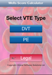
You’ll notice, it also has a calculator for PE risk, we will get to that more in the second part of this post series. The Wells App is not the only one, it just happens to use a criteria that is widely recognized. There are other options that utilize similar criteria like the Caprini. Either way, you should get one of these free apps on your device so you can perform in-the-moment assessments of your patients.

One of the toughest things is that, if you look at the Well’s score criteria, you will see that pretty much any and every total joint replacement patient is automatically at high risk. This is actually 100% true! We know this and this is the reason we educate patients regarding signs and symptoms of DVT, so they can be on the lookout for complications when we are not there to help them! However, that doesn’t mean you need to call the physician. Like I said above, you have to correlate clinically. If their Well’s score is high risk AND they’ve had a significant functional decline AND they have an extremity that is three times the size of the contralateral one, then you should probably be making a call.


Education
Patients get pretty worked up about risks after surgery and need a good amount of post-operative education and education reinforcement to help them make appropriate decisions about the possible presence of a DVT, so you want to make sure they know what to look for. One of my favorite things to tell them is how to differentiate between post-operative edema and DVT-related venous pooling is that the affected extremity (which MAY NOT BE THE OPERATIVE ONE!!!) will be two to three times the size of the other side, not just at the joint, but the entire extremity. This paints a pretty clear picture and the patient can actually be reassured that, because they do not look like this, they probably don’t have a DVT to worry about. However…

“…about 30–40% of cases go unnoticed, since they don’t have typical symptoms”

NIH

So we have to continue to be vigilant. We have the opportunity to see patients far more often in the sub-acute setting, especially home health care, than most other providers, so we have to have eyes on and communicate with our team.

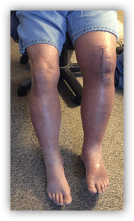
In my practice, I have identified several DVTs. The photo above of the positive DVT on the post-operative total knee arthroplasty was a patient of mine. My PTA saw this patient the day prior and called me to report something being wrong with his presentation. She asked me to see him for follow up first thing in the morning. At the time, he didn’t have the edema he has in the photograph. But it shows you that less than 24 hours can make a significant difference just in the physical appearance. He also had several other symptoms including confusion and a decreased functional status.

I have also identified DVTs in patients who were not post-operative, but were immobilized due to cancer treatment. It is important to keep in mind that, due to the cognitive symptoms patients can experience, even patients who are familiar with medical complications (such as doctors and nurses) can also experience DVTs if the conditions are right. So trust your clinical judgement, your gestalt feeling, and your evidence-based tools. Yes, I sent a doctor to the emergency department because I thought he had a DVT. He did. Like the ASH states:

“Blood clots do not
discriminate by age,
gender, or race. They
can affect ANYONE.”

ASH, 2018

COVID-19 and DVTs?


We know that patients with COVID-19 are definitely at higher risk for several reasons and through interference of several mechanisms (we talk about this here). Many of them are immobilized long term, whether in the hospital or at home self-isolating. A higher percentage of patients with COVID-19 have comorbidities like heart failure or diabetes that increase their general risk for DVTs. Newer studies are finding that the risk of DVT in patients with COVID-19 is about 20%. This strange thing called “blood stasis” in the femoral veins, which can lead to DVTs, is found in 70% of patients with COVID-19. That can be compared with the rate of DVT after joint replacement, which is about 0.15% for hip and 0.22% for knee – a massive difference.

And we also know that there may be an indirect mechanism by which this clotting occurs. When the endothelium is infected with COVID-19 and damaged, this triggers the clotting cascade, but this happens on a much larger scale than only a single point of compression or vascular insult. When the endothelium is damaged, it is damaged everywhere in the body that the viral RNA has infected it. When the patient has gone septic, that could be anywhere and everywhere. We talked about this in the post about how COVID-19 is recently becoming associated with increased risk fo CVAs.

Let’s also not forget that this risk persists long after discharge. We don’t know how long the risk persists. As long as the patient is recovering from COVID-19, whether it be in a rehab center or otherwise recovering with home-based therapy services, they continue to be at risk of developing a DVT or PE. According the ASH, 33% of people who have had a DVT will have another in less than 10 years time.

If you are already aware that your patient has a DVT, you are not technically excluded from exercising them. Let me be clear: if a patient has a NEW ONSET, acute DVT, you need to report this to the physician and get assessment and treatment immediately as risk of PE is very high. However, in patients who have acute DVTs under anticoagulation therapy or chronic DVTs (yes, these people are out there and you have probably seen them and not known it), you should be exercising them. According to the research, there is no increased risk for PE with exercise as compared to bed rest. Types of exercise included early mobility (household level) and ambulation, all the way up to vigorous treadmill programs (which actually showed fantastic long term results). If you think about it, these people are getting up and going to the bathroom and getting food aren’t they? So if household mobility does not increase risk, why would your interventions? Exercise should be implemented as soon as therapeutic levels of anticoagulation have been achieved.

You can review the Clinical Practice Guidelines for Physical Therapists in the Treatment of Patients with VTE:

Be sure to keep checking back for VTE Part 2: PEs! We will discuss how PEs are associated with COVID-19 and what you need to know as a rehab clinician.

What parts of the VTE CPG do you find surprising? What parts do you regularly implement in your practice? Let me know in the comments!

More Reads…

Pressure… Pushing Down On Me…

Breathing. I can’t stress it enough. If you’re not breathing, you’re dead… or in a lot of pain… either way, it’s not good. So breathe! In my practice, I work with a lot of different types of patients with a wide variety of conditions and comorbidities, but they all have one thing in common: they…

Dehydration

WHILE WE WAIT FOR THE NECT CHAPTER OF DIABETES MANAGEMENT, LET’S KEEP TALKING ABOUT INCONTINENCE

Chronic management of urinary incontinence can lead to many issues like infection and hospitalization if it doesn’t account for fluid balance! Let’s talk I’s and O’s! #physicaltherapy #incontinence #chronicdisease

Follow my blog for more!

Follow @DoctorBthePT on Twitter for regular updates!

References:

American Society of Hematology. (2018) ASH Clinical Practice Guidelines on Venous Thromboembolism. Retrieved from https://www.hematology.org/education/clinicians/guidelines-and-quality-care/clinical-practice-guidelines/venous-thromboembolism-guidelines on May 14, 2020.

Center for Disease Control and Prevention. (2020) Venous Thromboembolism (Blood Clots). Retrieved from https://www.cdc.gov/ncbddd/dvt/facts.html on May 14, 2020.

Hillegass, E., Puthoff, M., Frese, E. M., Thigpen, M., Sobush, D. C., & Auten, B. (2016). Role of physical therapists in the management of individuals at risk for or diagnosed with venous thromboembolism: evidence-based clinical practice guideline. Physical therapy96(2), 143-166.

Kahn, S. R., Shrier, I., Kearon, C. (2008) Physical activity in patients with deep venous thrombosis: A systematic review. Thrombosis Research. 122(6): 763-773. https://doi.org/10.1016/j.thromres.2007.10.011

Kerbikov, O. B., Orekhov, P. Y., Borskaya, E. N., and Nosenko, N. S. (2020) High incidene of venous thrombosis in patients with moderstae to severe COVID-19. medRxiv [Published ahead of review]. Retrieved from https://www.medrxiv.org/content/10.1101/2020.06.12.20129536v1.full.pdf

Lee, S. Y., Ro, d., Chung, C. Y., Lee, K. M., Kwon, S. S., Sung, K. H., & Park, M. S. (2015). Incidence of deep vein thrombosis after major lower limb orthopedic surgery: analysis of a nationwide claim registry. Yonsei medical journal56(1), 139–145. https://doi.org/10.3349/ymj.2015.56.1.139

Streiff, M.B., Agnelli, G., Connors, J.M. et al. (2016) Guidance for the treatment of deep vein thrombosis and pulmonary embolism. J Thromb Thrombolysis 41, 32–67. https://doi.org/10.1007/s11239-015-1317-0

image credit

So Now COVID-19 Results in Strokes (and other neat things about ACE2 receptors)

We can basically thank our old friend sepsis for the presentation of strokes in patients with COVID-19. This virus has changed so much and so frequently since it landed stateside, but there was evidence from its China days that neurological involvement was implicated. Initially, though, it all started in the lungs. We talked about this a lot over the last several posts, but ACE2 receptors, the primary receptors that have an affinity for the SARS-CoV2-19 virus, are present in the endothelium of the heart, arteries, kidneys, and in the glial cells in the brain. This is not new. I didn’t look very hard and found articles from 2005 that indicated that we’ve known this for a while. (Corona viruses are also not new, they just only really impacted animals prior to the SARS-CoV-1 and -2 outbreaks, so we’ve been researching this for a while.) Some evidence exists that suggests ACE2 receptors being present increase the risk of end-organ damage to that specific tissue because it receives the virus more easily. So here are some mechanisms by which COVID-19 can result in cerebrovascular accidents of different varieties.

image

Just a warning, we are going to get REAL SCIENCEY here!!! I love to tell my students to go back to the basics of what they know. Our basic physiology and anatomy knowledge is really important here. It’s important pretty much everywhere, but especially when discussing disease mechanisms. I know this stuff is tough but I’ve tried to make it straight forward. The better we understand our enemy, the more equipped we are to go in to battle.


Angiotensin converting enzyme 2 (ACE2) is on the outer surface of lung tissue which is why the lungs have been most heavily affected by COVID-19. We know the virus invades the lungs, overwhelms the natural protective factors due to expedited viral replication, destroys the lung parenchyma, and causes interstitial fluid build-up. Damage of the basement membranes of the alveolar pneumocytes results in scarring because the pneumocytes cannot restore themselves to their original location or function without the guide of the basement membrane.

MECHANISM 1. So what happens with ACE2 receptors in the heart? ACE2 receptors in the heart function to lower blood pressure via changes to stroke volume when they are activated. When these receptors are impaired or destroyed due to infection with the SARS-CoV-2 virus, blood pressure begins to elevate under little control. The more receptors damaged, the more blood pressure elevates. We know that when a hypertensive state is sustained or just very severe, a cerebrovascular accident of either ischemic or hemorrhagic etiology is possible. Then you pair this with ACE2’s function in the vasculature (vasoconstriction) and pressure increases even higher. If the patient has an underlying comorbidity (like A-Fib) that could result in clotting, ischemic etiology would be more likely as this pressure would dislodge a clot. If not, a hemorrhagic event could be suspected.

MECHANISM 2. How about the brain? ACE2 receptors are on glial cells but the blood-brain barrier is supposed to give us an extra level of protection against these types of infections. Unfortunately, the SARS-CoV-2 virus attacks the endothelium of capillaries and destroys this single-cell barrier (the blood-brain barrier) allowing the invasion of SARS-CoV-2 virus in to the neuroglia. These types of infections tend to result in cerebrovascular accidents of the hemorrhagic etiology. This can also result in peripheral nerve injuries to a lesser extent. It is suspected that this is also why people have chemosensory changes, confusion, and delirium as symptoms of COVID-19 before treatment.

Photo by meo on Pexels.com

MECHANISM 3. We know a patient with COVID-19 demonstrates increased blood viscosity, decreased perfusion, general ischemia/hypoxia, and requires increased cerebral blood flow to maintain function (because this blood is poorly oxygenated). As PaCO2 and PaO2 become altered, and the patient becomes hypercapneic, cerebral autoregulation which regulates intracranial pressure (ICP), is impaired. This state of increased pressure can also result in a hemorrhagic accident. Unfortunately vasopressors do not cross the blood-brain barrier so we cannot modify this via this mechanism and perfusion remains poor.

MECHANISM 4. COVID-19 viral RNA may attach to capillary endothelium when blood born (hello, again, Mr. Sepsis). We know this can result in a cytokine storm with end-organ damage targeting pretty much any system you can name. Included in this list of systems is the clotting cascade and endothelium themselves. This results in thrombosis and platelet activation which overall produces clotting. It is known that patients with COVID-19 develop pulmonary embolisms at an increased rate. This could be one of the reasons (along with immobilization, medications, etc). These clots can also go pretty much anywhere, including the heart, brain, kidneys, etc… More than 70% of people who perished due to COVID-19 demonstrated intramuscular thrombosis! This is NOT normal. So all these organs systems continue to be at risk for serious injury due to VTE risk. This risk continues after the ICU and rehab phases of this condition and persists with the patient. We don’t know how long yet. The risk of ischemic CVA increases with respiratory infection even in non-COVID-19 patients.


Here’s the really tough part. Some of the things we have to do to sustain life in a patient with COVID-19 may actually be causing cerebrovascular accidents or other neurological complications and extending the timeline of recovery. However, the risk/benefit analysis is pretty grim: definitely die now or maybe not die later. Our physicians and patients have really tough choices to make.

MECHANISM 5. A very high PEEP is required for mechanically ventilated patients with COVID-19, higher than most other diagnoses require. Positive End-Expiratory Pressure (PEEP), not to be confused with PEP, is pressure applied at the end of exhalation to maintain the patent airway. PEEP is typically seen in those who use CPAPs or BiPAPs. This must be more than the atmospheric pressure and more than the passive pressure of exhalation. Many articles I’ve seen talk about 5 cmH2O as a standard starting point for most patients. Patients with COVID-19 are requiring 30+ cmH2O to maintain their airways (I’ve read cases of 60+ cmH2O reported). The use of higher PEEPs results in increased ICP which can result (as we have already mentioned) in neurological complications and stroke.

Photo by Anna Shvets on Pexels.com

MECHANISM 6. Proning itself does not cause neurological complications, but it is difficult to maintain in most patients without the use of sedatives. If you think about the position with a giant tube through your trachea, lines and leads coming off every inch of you, and a generally uncomfortable hospital bed, you’d probably want sedatives, too. Sedatives can also be helpful for managing the discomfort that comes with sepsis. However, they can decrease cerebral perfusion and metabolism which further decreases cerebral autoregulation. If the patient is proned and positioned in a way that compromises the jugular vein, ICP can further be increased and we end up with similar issues as in mechanism 5.


Thankfully, ya’ll, I see a lot of room in here for rehab clinicians to intervene.

  1. TAKE VITALS – every patient every time, more than once, at rest and with activity, as often as is clinically indicated, and then maybe a few more! Monitoring blood pressure is going to be so important as hypertension is our biggest enemy here! Respiratory rates at rest and with activity, when combined with oxygen saturations can help determine presence of PE or DVT. Welcome to my soapbox.
  2. NEUROLOGICAL BASELINE ASSESSMENT – If you don’t remember what they looked like when they started with you, you have no basis for comparison if something changes! How confused were they? How delirious were they? Were they having headaches?
  3. POSITION SMARTProne smart. Position well. Change positions frequently. Create a turning schedule. Take charge of positioning schedules to ensure they are maintained. Changing patient positions helps reduce PE and DVT risk. Prevent peripheral nerve injuries and ICP elevation.
  4. MONITOR DRUG USAGE – Ensure patients are taking their medications as prescribed. Review their medications for changes or questions. Make sure they can access their medications and can get their refills when needed. Managing a patient’s medical comorbidities, like A-Fib, well reduces the risk of complications like neurological insult.
  5. ADVOCATE – in the ICU for early mobility and breaking or reducing sedation as early and as often as possible while maintaining a hemodynamically stable patient in conjunction with your interdisciplinary team. Reduce the risk of DVTs and PEs, reduce the effects of sedatives, and reduce the overall cognitive impact.

You guys are working miracles out there! Tell me how you and your team are addressing the risks of CVA for COVID-19 in the comments!

Home (Health) is Where the Heart Is

Dr. Rebekah Griffith, other wise known as The ED PT, interviewed Dr. B on all things patient care management and how PTs in the ED and Home Health PTs can work together to provide the best possible care for patients. We talked about the struggles we each face in managing complex patients and the roles…

Primary Care PT

PTs primarily treating diabetes shouldn’t be a far cry from the future, it should be today! Read this for more!

Something went wrong. Please refresh the page and/or try again.

Follow my blog for more!

Follow @DoctorBthePT on Twitter for regular updates!

References

Levenhagen, K., Gorman, S., Verma, A., Kumble, S., Lopker, M. (2020). Physical Therapy Considerations of Neurologic Presentations in COVID-19. [Webinar] Viewed May 8, 2020. https://register.gotowebinar.com/recording/8523389256235766031

Michigan Physical Therapy Association – Western District Webinar: Combating COVID-19

Doctor B was featured in a live webinar hosted by the Michigan Physical Therapy Association to provide clinicians ideas of how physical therapists should be combating the COVID-19 pandemic.

  • Options for interventions
  • Roles we can play
  • Health care system and supply concerns
  • Precautions
  • Physiology of COVID-19

View the Webinar Here!

Trouble loading? Here is the direct link: WEBINAR

A big thank you to the MPTA Western District for inviting me to provide this education to their members! Visit the MPTA website here!

image

Did you get a chance to watch the Webinar live? Let me know you were there in the comments!

More Reads…

Did Anyone Else See That?

Do you ever wonder, “did anyone else see that?” We find those red flags sometimes, don’t we? We see something odd in a physical exam and it completely changes our perspective on the patient. The key to seeing it, though, is to look for it. You may be thinking, “another thing I need to add…

More Than Just A Respiratory Disease: The COVID-19 Toolbox

Isn’t COVID-19 just a respiratory disease? If only that was true. We are good at treating respiratory infections. We have lots of drugs for viral, bacterial, parasitic, and fungal infections of the lungs. Most of them work really well! We also have several backup treatments, inhaled medications, and adjuvant therapies (like rehab!) that make primary…

COVID and Clotting: How to Identify, Assess, and Treat Clotting Disorders in COVID-19 Survivors

Treating patients with acute and chronic clotting conditions is not new to rehabilitation professionals. We even have clinical practice guidelines around how to do so safely. However, what is new is the increased risk of newly acquired clotting conditions among post-COVID-19 patients. Those of us practicing in acute, subacute, emergency, and outpatient settings are uniquely tasked with…

Something went wrong. Please refresh the page and/or try again.

Follow my blog for more!

Follow @DoctorBthePT on Twitter for regular updates!

PODCAST #1

Join Afrin Mandlik, PT and Doctor B as they discuss:


A Historical Perspective

How Physical Therapists Function in times of Pandemic, Epidemic, and Crisis.

Recorded March 24, 2020
(It’s amazing how much has already changed since then!)

Thanks so much for listening! I’d love to get your feed back!


Do you have requests for PodCast topics? Let me know in the comments!

More Reads…

Altitude Medicine: Rehab at the Peak

I took a trip to Denver recently and, right around the same time, was consulted regarding the use of a pulse oximeter at high altitudes. It all got me thinking: how different could it really be to practice at 9,000ft? So, I did some work on this and I have to tell you, finding this…

Emergency Response Screening

I hope, at this point, you’ve all heard of the FAST acronym for identifying signs of a cerebrovascular accident (aka stroke). My in-laws even have a magnet on their fridge with a great comic strip describing the FAST acronym. I have run in to a couple different versions of it in the last few years,…

Something went wrong. Please refresh the page and/or try again.

Follow my blog for more!

Follow @DoctorBthePT on Twitter for regular updates!

The Counting Talk Test

Time to give you guys another tool. This one is especially important for patients with COVID-19, and it’s not terribly often that we get an objective measure for exertion. I’ve found the counting talk test to be critical in quantifying exertion in my patients with respiratory diagnoses. This measure is phenomenal for taking breathlessness in to account.

Here’s how it works:

  1. At rest, have the patient take a breath and count out loud as high as they can. Record that last audible number. (FYI – this works best if done in standing, but be sure the patient is in the same position pretest and posttest)
  2. Have the patient perform the exercise you intent to prescribe.
  3. While they are working, have them perform the count again. Record the last audible number.
  4. Do some math using this worksheet

I know, I don’t like math either, but it’s simple math and it gives you a lot of information like amount of effort expended with exercise and precent workload which then corresponds to the Borg RPE scale. I created this worksheet with outpatient cardiac rehab or subacute rehab/home health in mind, so the workload goal at the bottom is a bit on the high end for a patient with COVID-19, where you should be targeting 4-5/10 at first.

Why do you need another scale for measuring exertion? You already have the Borg scale, the Borg RPE scale, the Borg Dyspnea scale, and the VAS for exertion… Here’s the thing: those are all subjective and are difficult to understand for people who have delirium, confusion, or cognitive impairment, as do many of the patients who have COVID-19 once they reach rehab. Some clinicians have found that, when working with patients with COVID-19, these factors prevent effective use of the typical RPE scales in practice. Yes, you also have blood pressure, heart rate, and oxygen saturation to rely on, but these things are affected by medications like beta blockers. So, although they are objective, they don’t give you a true clinical picture of the exertion the patient is experiencing.

You need something truly objective. Of course, if the patient has severe delirium, confusion, or cognitive impairment, they may not be able to count to 30. So you will have to rely on vitals, but since we know these are not a great representation, we should use the best tool we have if the patient is able to participate in it. This is a great test for the post-COVID population because exercise intensities are generally on the lower end so speech-rate variability with exercise does not impact the test (see link in references for more on this).

Most patients with respiratory conditions can count to some number in the high 20’s. I’ve had coworkers reach up in to the low 40’s during training with this tool! The cool thing is, it doesn’t matter what number they reach, because you retest every day and use their top resting number as your baseline for that session. We all know that people with pulmonary conditions look pretty different from one day to the next, so it’s important that the tool be as flexible as their presentations are.

This test is also great because it can be incorporated into ADL performance. We know that with many patients who have advanced pulmonary diseases or who have COVID-19, their tolerance to ADL can be significantly diminished. The counting talk test can also be utilized during ADL performance (like taking a shower!) to determine energy expenditures and RANK activities according to expenditure! This can guide interventions and point a clinician toward what activities need to be modified to decrease energy expenditure! Energy conservation interventions just got a whole lot more clearly identifiable! Obviously you don’t want to do several ADL in a row for measurement as the total workload would increase the energy expenditure. But at least this gives you a quantifiable way to document tasks.

When prescribing interventions, you can also use the energy expenditure numbers (as a percentage) to demonstrate improvement over time. If one minute of squats utilized 46% of my energy today, but after 8 weeks of boot camp, only used 34% of my energy, I could document evidence of cardiovascular reconditioning.


In the comments, tell me how you use the counting talk test in your practice!

More from the Pulmonary Rehab Toolbox…

Something went wrong. Please refresh the page and/or try again.

Follow my blog for more!

Follow @DoctorBthePT on Twitter for regular updates!

References

Validity of the CTT: https://www.ncbi.nlm.nih.gov/pubmed/18496320

Speech Rate Variability due to Exercise: https://www.ncbi.nlm.nih.gov/pubmed/30034101

Consistency of the CTT: https://www.ncbi.nlm.nih.gov/pubmed/21904237

Use in Cardiovascular Disease: https://www.ncbi.nlm.nih.gov/pubmed/25010379

Levenhagen, K., Gorman, S., Verma, A., Kumble, S., Lopker, M. (2020). Physical Therapy Considerations of Neurologic Presentations in COVID-19. [Webinar] Viewed May 8, 2020. https://register.gotowebinar.com/recording/8523389256235766031

What if this is what you did all day every day?

You walk in to work, you scrub in, gown, mask, shield, gloves, more gloves. You have to be let in to rooms so you aren’t contaminating your gloves. You have to be let out of rooms so you aren’t contaminating your gloves. You work in a negative pressure environment all day, every day. You wear an N95, PAPR, or N100 all day, every day.

Does that sound strange? Does it sound strange that you could possibly spend every day that you treat patients dressed in full PPE for the rest of your career? Does it sound strange that someone would actually choose that? Because that’s exactly what is happening in other countries. Physiotherapists from the UK to India are working their regular full-time days managing and treating patients who have tuberculosis.

Although we don’t see much TB here in the states, in other countries like England or India, or most everywhere else but here, TB isn’t uncommon. In 2018, TB killed 1.45 million people worldwide. That isn’t to say that there are tons of TB (+) people running about the streets, but there are enough to warrant special facilities for their care. It is at these special facilities that PTs and OTs jump in to action.

Patients who experience TB tend to have a long term presentation similar to COVID-19. They are hospitalized for a long time and usually end up very deconditioned. They need rehab to recover from PICS. They need cardiopulmonary PT interventions like using positive pressure devices to induce coughing and exercise to increase their strength. Previously thought to be unsafe, new guidelines are making this possible! They need ADL retraining and energy conservation education to return home safely. And they need to test (-) in order to be discharged home. There are some rehab clinicians who do this work as their full time job. You can read about them here!

Research has even shown that only eight weeks of pulmonary rehab involving all of these interventions makes significant (and large!) improvements in functional outcomes measures like the 6 minute walk test and in questionnaires like the SF36. You can read about that here!

Video credit: Sierra Stallard (ICU Nurse – Beaumont Royal Oak)

So, what is it like treating a highly infectious airborne pathogen day in and day out? Strenuous. Even though the NIH suggests a certain set of guidelines for care of patients with TB, most facilities implement the highest level of protections. That includes PAPR masks or other respirators, shields, gowns, gloves, more gloves and working negative pressure full isolation environments. Don’t forget anything you might need outside of that room! And the therapists can’t do it all day. They usually work half days in the facilities and half days elsewhere to prevent burnout and decrease viral load and exposure. And what about the physical effects of this work?

There has been some evidence that wearing respirator-type masks (some more than others) cause significant discomfort. We’ve seen pictures of our frontline providers with skin breakdown on their noses and cheeks from goggles and respirators, but that’s just the physical difficulties. There may actually be physiological impacts of wearing respirators long term including significant thermal stress to the skin and airway. This can lead to increased heart rate, dizziness, headaches, difficulty with decision-making… all the things we don’t want to be dealing with when we have major infectious agents to be thinking about. However, like I said, this research is preliminary. And it definitely does not apply to procedural masks or other face coverings which healthcare providers wear constantly.

Does COVID-19 still sound so terrible?

Working in a COVID-19 isolation unit still probably sounds pretty terrible. Working in an isolation environment all day every day is taxing. There is a significant increase in the amount of clinical decision making that has to take place when working in isolation with an airborne agent, especially when providing procedures and interventions that will definitely aerosolize the viral particles. Not to mention, there are treatments for TB that make exposure less of a risk, whereas COVID-19 still has no verified treatment, although it looks like some antivirals are promising. All of this clinical decision-making has to take place under possibly physiologically straining conditions.

However, like TB, COVID-19 knows no age limit, no comorbidity limit, no gender, and can present with neurological symptoms as well as cardiopulmonary symptoms. There are some schools of thought that are concerned that COVID-19 may even be airborne, like TB, due to the many healthcare workers that have contracted it regardless of environmental and PPE precautions in place.

Overall, COVID-19 is not the first time PTs and other rehab clinicians have been faced with treating highly virulent pathogens. It’s not the first time we’ve been asked to don full PPE before walking in to a patient’s room. COVID-19 is not the first time we have been asked to provide care under strenuous conditions. And it will not be the last time that we are asked to do any of these things. New pathogens emerge all the time. Tuberculosis persists. Providing the best rehab we possibly can will persist, too, all around the world.

We’ve got this. ❤

If you practice in a country other than the US, do you feel any different suiting up to treat COVID-19 than you do when treating any other communicable disease? Let me know in the comments!

Other Reads…

PT/INRs: Helping Patients Manage their Anticoagulation

So, who’s up for some bloodwork? Venipuncture, anyone? Finger prick, maybe? No..? Oh… Well, ok then. I’ll carry on. I know it may sound a bit crazy, but as a PT, I actually do some (very) minor blood work. I’m not trained as a nurse or phlebotomist or anything like that. Nope. Just some on-the-job-training…

It’s Getting Hot in Here: Body Temperature

How many times have you had your temperature taken lately? I think I’ve had my temperature taken thousands of times in the last few months. We are seeing the increased use of forehead scanning thermometers and temporal scanners, all the non-contact forms of temperature assessment, to screen folks for COVID-19 symptoms upon entry to any…

COVID-19: Where the Heck Are We?

I started this venture back in March in response to COVID-19. I saw many Physical Therapists and other rehab professionals shutting their doors because it wasn’t safe to keep them open. I saw an opportunity to improve our profession by addressing a deficit in practice: the lack of cardiopulmonary skill implementation. It wasn’t even that…

Something went wrong. Please refresh the page and/or try again.

Follow @DoctorBthePT on Twitter for regular updates!

Orthostasis

“Whoops! Down I go! I thought I was going up,
but apparently not!”


I have heard this many times. Sometimes I hear it from the people around me just generally throughout the day. They black out or see stars or feel “whoozy” or report being lightheaded when they stand up from the couch or get out of bed. I’ve seen people get up to do an OASIS walk, stumble, and bounce off a wall!

We’ve mentioned orthostasis several times and in great detail in my post about beta blockers. Patient goes up, blood pressure goes down (heart rate may or may not elevate depending on the patient’s ability to accommodate). Assessing this is a pretty integral part of my regular evaluation. However, I find that, although many clinicians easily recognize it, very few assess it, quantify it, and intervene against it. So why would you want to quantify it? So you can demonstrate improvement in the patient’s presentation and symptoms after you apply your interventions! Here’s what triggers this assessment in my evaluations:

  • Patient reports dizziness or lightheaded with moving around. Yes, this of course could be vertigo of some etiology. However, why not easily rule out what you can?
  • Patient had a recent cardiac procedure, especially an open heart procedure like a CABG or AVR. But even closed procedures like TAVR and cardioversions can result in new or reoccurring orthostasis.
  • A change in medications that effect blood pressure, whether it be a new addition, a dosage change, or a removal of a medication. Pretty much any cardiac or pulmonary medication change
  • Patient has been immobilized for an extended period of time, greater than 4 days, generally. So this would be really relevant for people who have recently been in the ICU.
image

What leads to orthostatic hypotension?

So many things. I mentioned above that any changes to cardiac and/or pulmonary medications can result in orthostasis. So an obvious one to consider here. But there are other less obvious causes that need to be considered. Blood pressure is a function of blood volume, so if you don’t have enough volume (hypovolemia), you won’t have enough pressure. This is largely an issue in patients with COVID-19 as they are kept intentionally “dry”. Some medications, such as hydrocholorothiazide (HCTZ), lasix (furosemide), aldactone, polyethylene glycol (miralax) and many other medications like them literally pull water out of your body and in to other systems like the kidneys or the colon to be excreted.

There are also dietary factors that contribute to dehydration such as just basically not drinking enough water (I promise you this is not as obvious as you think), ingestion of diuretic items, such as coffee, and a high-salt diet. Patients who have a fluid restriction, such as those with End Stage Renal Disease (ESRD) or congestive heart failure, can actually be afraid of taking in too much fluid because it could exacerbate their disease state. This can result in dehydration due to not taking in enough fluid to function at the basic physiological level.

Blood volume is also affected by hydration status. Blood is about 92% water! You may be thinking, “uh, my patient isn’t bleeding out so I’m pretty sure they have enough volume.” Don’t jump so quickly to that conclusion. Your patient may actually be bleeding. Occult GI bleeds are fairly common in the elderly and chronically ill populations. I’ve also seen several chronic subdural hematomas. Just because you can’t see it, doesn’t mean they aren’t bleeding.

If your patient is dehydrated, their blood volume may not be sufficient to support a functional blood pressure that can perfuse the brain when having to work upward against gravity. There are several ways to check for dehydration in most patients. The skin turgor test is a quick and easy way to know, but you also have to correlate it with clinical symptoms to increase its specificity.

Skin Turgor Test
image

You can also have a discussion with the patient (or observe for yourself) about their symptoms. Things such as:

  • headache
  • decreased urination
  • dark urine
  • thirst (however this is decreased in older people)
  • dizziness
  • nausea
  • falls (in more severe cases)
  • confusion (in more severe cases)
  • UTIs (in more severe cases)

All of these things contribute to

H e m o d y n a m i c S t a b i l i t y

which is one of my favorite things to talk about with clinicians, patients, and students. Applying the basics of what we know can so greatly inform our practice.


So what do we do about it

You can get all the details on assessing and quantifying orthostatic hypotension in my post about beta blockers. There is even a handout to print, some tips for reporting, and how this whole process works. But once you know it’s there, you need to do something about it. You have a few different avenues to explore…

  • If orthostasis is due to hemodynamic instability (low blood volume, dehydration):
    • Increase fluid intake. Increasing volume increases pressure.
      • Be aware of fluid restrictions for ESRD/CHF
      • If fluid intake cannot be increased, accommodate with compression stockings and/or abdominal binder.
    • Implement orthostatic support exercises
      • Perform arm cycling, ankle pumping, marching, etc, based on patient needs while in supine for 30-60 seconds prior to sitting.
      • Perform arm cycling, seated LAQ, seated marching, etc, based on patient need while in seated for 30-60 seconds prior to standing.
      • If still symptomatic, stand in place until symptoms clear before ambulating to reduce fall risk.
  • If orthostasis is due to long term positioning, vestibular accommodation, or neurological disorder (such as Parkinson’s Disease):
    • Utilize slow progressions between position changes. Consider use of a tilt table.
    • Compression garments such as stockings and abdominal binder may be helpful.
    • Consider use of pharmacological interventions to elevate blood pressure.
  • If orthostasis is due to medications:
    • Contact the physician to report your findings and ask if there are any changes they would like to make. It is very common that patients tolerate these medications at first or don’t get up as often after procedures that necessitate them, so symptoms are not spotted early on. You may be finding this later, but that doesn’t mean anyone else knows about it.
    • In the mean time, implement orthostatic support exercises as noted above.

Don’t Forget…

When assessing orthostatic hypotension, record blood pressures, heart rates, oxygen saturation (if able), and patient symptoms in supine, seated, and standing and report ALL of these things to the physician. If blood pressure or heart rate goes outside of your physician-, agency-, or facility-established protocol at any point, notify the physician.

If the patient’s blood pressure or heart rate goes below the bottom line acceptable value at any position, stop the assessment. These numbers may be patient specific, pre-established, or physiologically determined. Typically, if I’m getting numbers in the 70s over 40s for blood pressure, I’m stopping the test and getting the patient in recovery position immediately, even if they are not symptomatic. This blood pressure is not sufficient to perfuse the brain. Risk of falls becomes very high. The standard cutoff is 90/60 mmHg if the patients has no other set values, especially if they are symptomatic.

And once you have checked all of this, reported what needs to be reported, and applied interventions, don’t forget to RETEST!!! You can document some remarkable improvements in patient function and reduced fall risk all because you decided to look beyond the surface.


Well, that was fun! What exercises do you like to use to prevent orthostasis between position changes? Let me know in the comments!

More from the Pulmonary Rehab Toolbox…

CO2 Retainers

MORE BONUS CONTENT! Getting sciencey now! The science behind COPD and oxygen to help lead you right!

Follow my blog for more!

Follow @DoctorBthePT on Twitter for regular updates!

Call in a Consult

If you know me or if you’ve ever worked with me, you will know that I am a huge fan of multi-disciplinary care. I am always trying to look for underlying issues that can be addressed by my colleagues and teammates that can improve outcomes for my patient beyond what I can provide them. COVID-19 is no different.

There are many side effects of not only the pulmonary condition itself, but the resulting conditions of acute respiratory distress syndrome and post intensive care syndrome that not only warrant, but mandate a multidisciplinary approach.

I’m feeling a bit salty today, so I’m going to be implementing some memes
for my own entertainment.

I don’t know if you’ve ever been intubated, but I have. For a procedure that would take longer to write out than the 45 minutes the procedure actually lasted, I was intubated. I needed an upper airway reconstruction of sorts because I had been diagnosed with obstructive sleep apnea when I was 21 years old. Turns out I was pretty anatomically dysfunctional, and my airways were just not performing the way they should have. So, after eight months on a CPAP which I completely failed, we decided to go forward with the surgery.

I’m betting you’ve had at least one patient who uses CPAP. Do they tell you that it’s extremely uncomfortable and they don’t like wearing it? They are 100% correct! CPAPs are incredibly uncomfortable and difficult to wear if it’s something you’re not used to. I’ve talked with many people who use them religiously and could never sleep without them, but in my personal experience, it was some of the most uncomfortable nights I’ve ever had. It was hard to breathe at all with that pressure blowing back in my face. I utilized a nose-mouth mask, but quickly had issues with claustrophobia, so switched to a nasal mask only. I attempted to use nasal pillows, but couldn’t keep those on any better than I could the regular nasal mask.

I cannot tell you how many consultations I had with my respiratory therapist, Holly, who is one of the most amazing people I’ve ever met. Holly did absolutely everything she could and gave me every mask she could find to figure out one that worked for me. She was in constant contact with my ENT to adjust pressure settings to improve my comfort and tolerance while still maintaining patent airways. Eventually, Holly and I decided that, given my age and my intolerance to the device, it was time to move to surgery. And she was right! The surgery was successful and I’ve never had a problem sense.

I bring this up because, even though I had seen a respiratory therapist for my initial mask fitting, I had always gone back to my ENT to address my intolerance of the device. I was still in graduate school at the time, and had not had a large amount of exposure to respiratory therapy other than my own personal experience. Had I known that consulting my RT was the way to go from the start, I could’ve save myself a lot of discomfort and time and gotten much better sleep. Inter-disciplinary consults work the same way for COVID-19.

Thankfully, respiratory therapists are already integrally involved in the treatment of COVID-19, so they will likely be the ones consulting us, not the other way around. But there are some other disciplines we may want to consider. Like I said, I’ve been intubated in the past, but only for 45 minutes! And even after that very short amount of time, I still experienced some problems. My ENT had warned me that I would be a challenging intubation because I have very small airways, and a shorter neck (in his words…) Can you imagine how patients with COVID-19 feel after days worth of intubation??? Not great, I’m betting…

So, after I came out of surgery, my neck was killing me. Two days later I saw physical therapist for some cervical spine work and one visit was enough to correct the issues and I had no further problems. Although we may initially think of physical therapy being utilized for COVID-19 to treat post-intensive care syndrome and impairments to airway clearance and diffusion, our orthopedic skills can still be just as important.


I’ve had many patients that were intubated for heart and lung transplants. Some of these surgeries last 14 hours and the patient is intubated for the entirety of the procedure. Afterwards, their swallows is so impaired that they end up on tube feedings. Their vocal cords can be damaged creating vocal and volume impairments. They can also have severe throat soreness. All of these are things that can be addressed by consults to our wonderful friends the speech therapists.

If you have a patient in any setting who is been discharged from the hospital for any reason and is experiencing any of these symptoms after being intubated, they need a consult from the speech therapist. The symptoms can be short-lived, but they can also be very persistent. Most of my patients did end up getting off of the feeding tube after transplant, but some did not. If you read my posting about acute respiratory distress syndrome and the other posting about post intensive care syndrome, you will have learned that there are significant cognitive and mental health side effects of both of these conditions. Possible delirium and cognitive impairment are things that need to be evaluated for baseline function and measured for return to normal by our speech therapist associates. And there’s that possibility of a feeding tube after being intubated, so the SLP is so crucial in returning the swallowing capabilities of our patients!

After patients leave the hospital, they may be coming home on a BiPAP, a CPAP, or possibly even home ventilation. If this is the case, there’s a lot of education that physical, occupational, and speech therapist can provide, but if you have access to a home care-based or outpatient respiratory therapist to help with management of all of these new devices, this patient will need direct support.


And then there’s our close colleagues the occupational therapists! Many of these respiratory devices come with masks that are difficult to manage and take a significant amount of strength to don and doff. The range of motion required to get a mask overhead is sometimes more than a patient is able to muster, especially after experiencing post intensive care syndrome. We’ve learned more recently that COVID-19 affects the smaller muscles of the body, so we can expect that the hands, arms, wrists, and fingers will be affected significantly. Velcro closures are challenging enough to figure out what goes where on these masks, but trying to manage them when you have no strength it’s going to be so much more difficult.

And that’s just the pulmonary device aspect of coming home! These patients will still need significant education on energy conservation, probably to the point of rearranging large portions of their home, so that they can still continue to function in their own environment without exhausting themselves. I once had to teach my mother how to put on deodorant with one arm after she had a wrist surgery. She had no idea that this was possible! Most people don’t. But our occupational therapists can come in and teach our patients how they can do more things with less.


If you have access to dietary services, a dietician is going to be an integral part of your team for patients after COVID-19. We typically think of modifying our diet to promote wound healing, but the wounds that COVID-19 patients experience are all on the inside. Their lungs have experienced a lot of damage and it’s going to take a change in their caloric intake to support healing from this damage. They also need to be taking in enough calories to support their activity levels which are going to be significantly increasing now that they’ve gone from being bedbound on a ventilator to having several therapies every day. And do you remember that feeding tube thing I mentioned earlier from being intubated for a while? Our dietitian friends can help with this, too! We also know that many of the patients who have had COVID-19 and need our care will also have several comorbidities that need to be addressed.

Many of these comorbidities have dietary restrictions such as sodium limitations, fluid intake restrictions, and different food group exceptions. Did you know a renal diet needs to be very low and beans? And since we know that 30% of patients who have COVID-19 in the ICU end up with renal failure and on a hemodialysis program, this information is important for their recovery. Many of them also have diabetes and need to maintain very strong blood glucose control to promote healing in their body. Our dietitian friends can help with all of these things.


And because most of them are going to be coming home at a level that’s probably not what we would consider ideal, they’re going to need access to as many community resources and services as we can fine for them. Because of the emotional and mental health inferences of these experiences, they may also need access to support groups and counseling. This is a perfect opportunity for our social work colleagues to step in and play a role. The social worker can work directly with the case manager at the physicians office, or with friends and family of the patient that they may not be able to contact due to continued isolation after returning home. They can figure out how to navigate some of the financial burdens many people are now experiencing and what assistance may be available, financially, emotionally, and physically. It may also be the case that these patients are returning home on hospice care. A social worker needs to be involved to ensure end of life measures have been properly established within the patient’s wishes.

And this is all assuming our nurse friends are already involved in managing all of the new medications, changes in medications, new medical diagnoses, and all the inter-related system presentations that we are all going to be struggling through to get our patient back to optimal health. Like I said in another post: Teamwork makes the dream work! 🙂


Do you have a favorite story working with an interprofessional team? What disciplines were represented? Let me know int he comments!

More Reads…

Sternal Precautions

“Patients exchanging habits of activity for complete rest are likely to become rapidly worse.” This quote fuels my everyday. These words have informed nonsurgical and surgical rehabilitation and its evolution from handing out bed rest like Oprah hands out cars to getting people moving early and keeping them moving often. Here’s the crazy thing: This…

Diaphragmatic Breathing

Let’s talk about this super simple technique that can change everything. Diaphragmatic breathing is really just how we are all supposed to be breathing most of the time. The purpose of the diaphragm is to facilitate breathing. Diaphragmatic breathing improves gas exchange and increases lung volumes. These are all really good things if we need to…

I’ll Huff and I’ll Puff… But the Huff Will Be More Effective

What is a huff? Huffing is one of the more difficulty techniques to learn for airway clearance. I find that most of my patients have trouble mustering the strength to perform a good solid huff. It’s not just that it is unfamiliar, but also that it takes diaphragm strength that they just don’t have. But…

Something went wrong. Please refresh the page and/or try again.

Follow my blog for more!

Follow @DoctorBthePT on Twitter for regular updates!

image credit

The Beta Blockade

Ugh, medications… I know, I know. I will try to make this as painless as possible. This one matters. It REALLY matters. For patients who have COVID-19 and for patients who don’t. Please take a quick read. Let’s start off by asking some questions.


What does a β-blocker do?

  • Blocks norepinephrine and epinephrine from binding to beta receptor sites (β1, β2, and β3 receptor sites), increases parasympathetic responses by blocking sympathetic input
  • Primary uses: Reduce heart rate, reduce blood pressure, vessel dilation, regulation of abnormal heart rhythm, reduce myocardial oxygen demand.
    • Overall decrease in mortality and morbidity.
    • Decrease abnormal ventricular remodeling of cardiac muscle tissue due to disease states.
  • Adverse effects: airway constriction, sudden heart failure, shortness of breath (in asthmatics), masking of low blood glucose symptoms in diabetics
  • SAFETY LIMIT: MediSpan and other medication management tools limit medication that effect blood pressure to four per patient. If this number is exceeded, risk of orthostasis is significant. Beta blockers would be included in this limit.

β-receptors? Can someone remind me what those are for?

Why sure!
β1 receptors on the heart increase heart rate, heart contractility, and cardiac muscle conductivity. These are cardioselective receptors so you won’t find them elsewhere.
β2 receptors are in the lungs and smooth muscle of the vasculature. They increase bronchodilation and vasodilation.
β3 regulates catacholamine-induced thermogenesis. (Part of how we create heat)

image

What are common β-blockers?:

  • Atenolol (Tenormin)
  • Acebutolol (Sectral)
  • Metoprolol (Toprol)
  • Propanolol (Inderal)
  • Carteolol (Cartrol)
  • Timolol (opthalmic use)
  • Sotalol (Betapace)
  • Labetalol (Trandate)
  • Carvedilol (Coreg) (β1 and α1 receptor)

Be on the lookout for duplicate therapies!
Beta-Blockers are often administered orally, but are sometimes administered opthalmically or by nebulizer (β2 receptors are in the lungs, remember!). Patients who are on Coreg or Labetalol as well as another Beta-Blocker (usually metoprolol) have a higher likelihood of orthostasis.

Patients who utilize beta blockers typically do so immediately after a cardiac procedure or to regulate chronic atrial fibrillation (A-Fib). There are other abnormal heart rhythms that beta blockers can be used for, but A-Fib is the most common. If you have a patient who has had an ischemic stroke, you can bet money on them taking beta-blockers. Irregular heart rhythms like A-Fib increase risk of clot formation and, therefore, ischemic strokes.


So if we have a patient on beta blockers, what do we need to do?

Monitor for adverse events. With orthostasis being so risky for our older population and so common when present in addition to dehydration, you have to be on the look out for it all the time. We’ve talked about orthostasis before and identified it as a drop in blood pressure due to changes in position against gravity. Some days it may be present and some days it won’t be. So what do you do if you expect it? Well, you need to assess orthostatic vitals. The CDC has a basic assessment handout for this. I’ve added it here for you to download (see download below picture). (Source) It is part of the STEADI toolkit, if you are a geriatric provider and are familiar with that.

So here’s the rub. (Ha! Cardiac joke…) My patients tend to be very low level, acute or chronically ill, on many medications, and/or unable to safely stand due to hypotension. So, I ask my patient to transition from supine to seated FIRST, wait there for 1 minute while I take their blood pressure, and then progress to standing IF they are at least 90/60 or not terribly symptomatic. However, I cannot tell you how many times I haven’t been able to let someone stand because they’ve already dropped below 90/60 in sitting. Please, PLEASE add a seated phase to this assessment.

Educate patient on signs and symptoms of orthostasis so they know when they need to sit or perform some activity in seated prior to standing to increase their circulation and blood pressure prior to standing. And when you do exercise them, there are a few factors you need to be thinking about.


So why do patients with COVID-19 need beta-blockers?

Let’s remember some important things about COVID-19. It is a viral infection that, if it damages enough alveolar basement membranes and parenchymal cells, creates interstitial edema which results in ARDS. This damage is severe and requires some type of ventilatory assistance to sustain life. Some patients can recover from this as their bodies heal the damaged pneumocytes with either scar tissue (which doesn’t diffuse oxygen) or replacement parenchyma (if the basement membranes were not destroyed). Either way, they will be in a significantly oxygen-deficient state for some time.

We know that these patients don’t typically go home after ICU. Sometimes they go to sub-acute settings like rehab or skilled nursing, and other times they transition to home because it wouldn’t be safe for them to be in a group setting. But overall, they need more time to recover than what acute care, understandably, has to offer. “When COVID Comes Home” is something I’ve focused on here for over a month. This sub-acute time frame is now being recognized as the next “COVID nightmare”.

So when these patients come home or head to rehab in this deoxygenated state, they don’t magically get better with supplemental oxygen. They still have significant oxygen diffusion problems due to damaged lung tissue and we, as the rehab clinicians, are about to ask them to start working a bit harder. It is estimated at this point that this may las tup to two months after extubation.

Then we add some comorbidities into the mix. We know that people who have an already compromised cardiopulmonary system are more likely to contract COVID-19. Our patients with COPD, Diabetes, Heart Failure, history of AMI… the list goes on. These patients are already on beta blockers in addition to a host of other medications that also have effects on heart rate and blood pressure.

Why? Why is this the case? If you remember our talk in my post on pulse oximetry, I discussed how to tell the difference between deconditioning, orthostasis, and diffusion impairment based on vitals response to activity. When patients with diffusion impairments exercise, their O2 saturation drops. In an effort to compensate for this, the heart rate and blood pressure will rapidly rise and the patient will very quickly lose their exercise tolerance and/or cross the barrier into unsafe vitals response to exercise. We need to temper the quick desaturation with beta blockers to reduce cardiac oxygen demand so that the cardiac muscle tissue is less affected by ischemia. Beta-blockers are the answer to prevention of this entire compensatory response that will allow vitals to remain within tolerable range longer to support functional mobility, and eventually exercise and rehab.


What do I need to know for exercising these patients?

People who are on beta blockers experience a blunted heart rate response to exercise. What that means is that the beta blocker medication is working to decrease cardiac oxygen demand by decreasing heart rate. It will take your body longer (about 5 minutes, sometimes more) to overcome this mechanism and begin to elevate heart rate in response to exercise (aka increased oxygen demand). So you need a warmup. And not just a few stretches. You need a progressive warmup that slowly increases oxygen demand, ramping upward toward your target intensity of activity over the course of 5-10 minutes. This also means that, while you are exercising them, you cannot use heart rate as a measure of exertion. You have to use something else like an RPE scale of some sort, or maybe the RPD Scale.


What activities do you like to use to prevent orthostasis in your patients? Let me know in the comments!

More from the Pulmonary Rehab Toolbox…

Blow Out the Candles…

If there is any treatment that I feel like gets used in a cookie-cutter fashion, it’s pursed-lip breathing. As much as I hate seeing this technique used for every single patient that has shortness of breath, it does have clinical usefulness. So let’s talk about how to implement pursed-lip breathing properly based on patient presentation…

Postural Drainage

We’ve all seen that dreaded picture in our textbooks… All the human figures laying in so many different positions with pillows and tables tilted all over… and I very clearly remember thinking, “How on earth am I supposed to remember all of those?” Well, good news. You really don’t have to. It’s great if you…

Something went wrong. Please refresh the page and/or try again.

Follow my blog for more!

Follow @DoctorBthePT on Twitter for regular updates!

References:

Hillegass, E. (2020). Vital Signs, oxygen, & exercise prescription: How are these impacted by COVID-19? PACER Project. Cardiovascular and Pulmonary sections of the APTA. Retrieved from https://www.youtube.com/watch?v=lj716KWNcig

Proning

We have been hearing all kinds of things about proning. What’s the big deal? Aren’t we just flipping people over? Well… yes! That’s exactly what we are doing. But here’s why its a bit more complicated than that.

We’ve already talked about the role of PTs in supporting and improving V/Q matching. Proning helps with that in a different way than what we’ve previously discussed. Basically what you need to know is that the lungs work on a negative pressure system. The pressure balance is a bit fragile and the pressure inside of the lungs must stay higher than the pressure on the outside of the lungs for the lungs to stay open. This is true not just for the lungs as a whole organ, but also for each tiny alveolus.

If the pressure on the inside of this tiny pocket becomes less than the pressure pushing in from the outside, the alveolus collapses. This can happen with destruction from ARDS, but it can also happen temporarily from laying in one position too long or from atelectasis or consolidation. The external pressure of the surface you are laying on slowly compresses the lung tissue in the dependent position (on the bottom of the body).

Proning allows for increased V/Q matching as less lung tissue is compressed in the prone position. The heart is located anteriorly, so there is less total lung tissue anteriorly than posteriorly. If we reduce the compression of the posterior segments, we improve expansion of the majority of the lung tissue and, therefore, improved oxygenation.

Just flipping over may seem really simple and obvious. But we need to consider the whole patient. Patients in the ICU are supine with a ventilator, multiple lines, multiple tubes, a catheter, and so many other things. These all have to be managed during the proning process. However, the results can be really great, especially in patients with COVID-19.

Photo by cottonbro on Pexels.com

Recent research has show that proning significantly improves mortality in patients with COVID-19 who have been intubated due to ARDS and are in the ICU. And I’m not talking, reduced by a little. I’m talking CUT IT IN HALF! Mortality from ARDS in the ICU at 24 days was reduced from 33% to 16% and at 90 days was reduced from 41% to 24%! This is no joke. This is a serious factor in improving outcomes for the most critically ill patients.

I’ve compiled some information from a video and three articles about proning to write this article. I am NOT part of a proning team, but this is just such an intriguing topic that I had to know more! So, I wanted to share with you what I learned. None of this is from my own experience except for where PTs can fit in to this team.

Now, about that team… You will need a few things set up and you will need to know the contraindications before getting started.

The Set-Up:

  • All necessary lines and leads need to be in place and secured
    • Arterial line
    • Central line
    • Lateral chest tubes disconnected and secured
    • Drains secured with abdominal binder with bulbs on the back
    • Feeding tubes disconnected
    • Hemodialysis lines disconnected and secured
  • All lines above the waist need to have their connected devices above the head of the bed and all lines below the waist need to have their connected devices below the foot of the bed.
  • Protective dressings in place over boney prominences
  • Patient has been suctioned recently
  • EKG leads removed (they will be replaced on the back later)
  • Patient’s gown removed
  • FiO2 at 100%

Your team will need:

  • Mepilex dressings
  • Tubegrip/Tape/splinting material to secure lines/leads/tubing/catheters
  • 3 bed sheets
  • 6 pillows
  • (Ideally) 7 people (1 RT/Pulmonologist/Very competent physician) and 6 of your closest friends. This can be done with fewer people but it makes it a bit more challenging.

Contraindications

  • Anterior chest tube
  • Hemodynamic instability
  • Facial/Ocular Injuries
  • Recent Sternotomy
  • Abdominal Incisions
  • Unstable Vertebral fractures
  • Unstable Intracranial Pressure that is Unmonitored

Here’s a great video of what this process looks like! The proning process starts at 7:30. Feel free to fast-forward.

Look scary? YEP! That’s ok. You’re not alone. An RT typically leads the show because they have to maintain the endotracheal tube placement throughout the movement. Watch some videos, including the AMAZING one I have above and get some ideas of how this goes and what you can do.

Here’s some great parts for PTs to play in this process:

  • Cervical spine stabilization: During proning, the cervical spine should not extend due to central line placement. PTs can assist in proning by stabilizing the cervical spine. Think of creating a cervical spine collar with your arms, from head to thoracic spine, to provide complete stabilization. Cervical spine positioning is also important to ensure proper management of intracranial pressure.
  • Code response team: If you find a patient pulseless while prone, you have to start CPR. But how the heck do you do CPR when you can’t flip the patient on your own? According to ACLS, you do compression to the same rate and depth over the thoracic spine at the level of the inferior angle of the scapula. This continues as typical CPR until the proning team can supine the patient and continue standard CPR. (PLEASE REMEMBER: There is no such thing as an emergency in a pandemic. Protect yourself and don all proper PPE before entering the room to assist in a code.)
  • Be a Roller: A roller is one of the 6 people on the proning team who roll the patient from supine to prone or back. Human movement is literally our job, so let’s do it.
  • Line and lead arrangement: We do this anyway, right? We go in to see a patient and those things are just all over the place. Even in my settings, I’m working with Foleys, ports, and IV lines for antibiotics pretty much every day. We know how to arrange this to best serve the mobility task we are looking to perform.
  • Positioning Post-Proning: Just like any other positioning program for off-loading, these proned patients need regular changes of position of many body parts every 2 hours. This includes utilizing “swimmer’s position,” head turns, and adding or removing pillows. Believe it or not, head turns require the full proning team!!! Peripheral nerve injuries can occur when patients are proned, so repositioning needs to happen frequently.

We can do this. We can take on this role. It is hard, clearly, as it takes 7 people to pull this off successfully, but you can be part of a really cool process that is nearly completely unique to what we have seen in the past and how we have functioned as a profession.


Are you part of a proning team? What are some ways you do this differently or have found that make it easier? Tell me about it in the comments!

More from the Pulmonary Rehab Toolbox…

Aerosol Generating Procedures

The long awaited clarification on aerosol generating procedures for physical therapists and physical therapist assistants has finally dropped! The APTA just released its professional guidelines for what portions of physical therapist and physical therapist assistant care equates to an aerosol generating procedures, therefore requiring increased PPE for procedure performance to ensure clinician safety. On April…

The Duet Device

Have you seen an Acapella Duet before? This is another one of those things I wish I could hand out to maybe half of my patients. A Duet device is a special kind of Positive Expiratory Pressure (PEP) that provides resistive oscillatory pressure to exhalation which promotes increased lung volumes due to re-inflation of collapsed…

FEV1

I spent some really great times as an educator in a heart and lung transplant program at a large hospital system. Specifically, I was a therapy educator. I taught PTs, PTAs, OTs, COTAs, and SLPs what they needed to know to safely provide rehab to patient after heart and lung transplants. I saw so many…

References

Ali, H. S., & Kamble, M. (2019). Prone positioning in ARDS: physiology, evidence and challenges. Qatar Medical Journal2019(2), 14. https://doi.org/10.5339/qmj.2019.qccc.14

Henderson, W. R., Griesdale, D. E., Dominelli, P., & Ronco, J. J. (2014). Does prone positioning improve oxygenation and reduce mortality in patients with acute respiratory distress syndrome?. Canadian respiratory journal21(4), 213–215. https://doi.org/10.1155/2014/472136

Lee, J., Kim, E., Song, I., Jung, S. A., Oh, H., Kim, J. (2016). Optimizing Prone CPR: Identifying the Vertebral Level Correlating With the Largest LV Cross Sectional Area Via CT Scan (Abstract for Presentation) American Society of Anesthesiology: Anesthesiology Annual Meeting. Retrieved from http://www.asaabstracts.com/strands/asaabstracts/abstract.htm?year=2016&index=4&absnum=4079.

Mount Sinai Health System Proning Team. (2020). Prone Positioning for the COVID-19 Patient. Retrieved from https://www.youtube.com/watch?v=ECdxhNFLwVo

Follow @DoctorBthePT on Twitter for regular updates!